 Movie
Movie Controller

[English] 日本語
 Yorodumi
- PDB-4dec: Crystal structure of glucosyl-3-phosphoglycerate synthase from My... -
+ Open data

- Basic information
| Entry | Database: PDB / ID: 4dec | ||||||
|---|---|---|---|---|---|---|---|
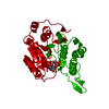
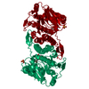
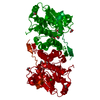
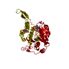
| Title | Crystal structure of glucosyl-3-phosphoglycerate synthase from Mycobacterium tuberculosis in complex with Mn2+, uridine-diphosphate (UDP) and phosphoglyceric acid (PGA) | ||||||
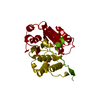
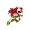
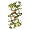
 Components Components | GLUCOSYL-3-PHOSPHOGLYCERATE SYNTHASE (GpgS) | ||||||
 Keywords Keywords | TRANSFERASE | ||||||
| Function / homology |  Function and homology information Function and homology informationglucosyl-3-phosphoglycerate synthase / UDP-alpha-D-glucose metabolic process / hexosyltransferase activity / glycosyltransferase activity / magnesium ion binding / protein homodimerization activity / metal ion binding Similarity search - Function | ||||||
| Biological species |   Mycobacterium tuberculosis (bacteria) Mycobacterium tuberculosis (bacteria) | ||||||
| Method |  X-RAY DIFFRACTION / SYNCHROTRON / MOLECULAR REPLACEMENT / molecular replacement / Resolution: 1.98 Å X-RAY DIFFRACTION / SYNCHROTRON / MOLECULAR REPLACEMENT / molecular replacement / Resolution: 1.98 Å | ||||||
 Authors Authors | Albesa-Jove, D. / Urresti, S. / van der Woerd, M. / Guerin, M.E. | ||||||
 Citation Citation | Journal: J.Biol.Chem. / Year: 2012 Title: Mechanistic insights into the retaining glucosyl-3-phosphoglycerate synthase from mycobacteria. Authors: Urresti, S. / Albesa-Jove, D. / Schaeffer, F. / Pham, H.T. / Kaur, D. / Gest, P. / van der Woerd, M.J. / Carreras-Gonzalez, A. / Lopez-Fernandez, S. / Alzari, P.M. / Brennan, P.J. / Jackson, M. / Guerin, M.E. | ||||||
| History |
|
- Structure visualization
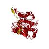
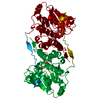
| Structure viewer | Molecule: MolmilJmol/JSmol |
|---|
- Downloads & links
-Download
| PDBx/mmCIF format | 4dec.cif.gz | 77.4 KB | Display | PDBx/mmCIF format |
|---|---|---|---|---|
| PDB format | pdb4dec.ent.gz | 55.5 KB | Display | PDB format |
| PDBx/mmJSON format | 4dec.json.gz | Tree view | PDBx/mmJSON format | |
| Others |  Other downloads Other downloads |
-Validation report
| Arichive directory | https://data.pdbj.org/pub/pdb/validation_reports/de/4decftp://data.pdbj.org/pub/pdb/validation_reports/de/4dec | HTTPS FTP |
|---|
-Related structure data

-Links
 PDBj
PDBj


- Assembly
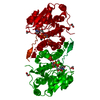
| Deposited unit | 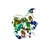
| ||||||||
|---|---|---|---|---|---|---|---|---|---|
| 1 | 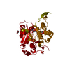
| ||||||||
| Unit cell |
| ||||||||
| Details | biological unit is the same as asym. |
-Components
-Protein , 1 types, 1 molecules A
| #1: Protein | Mass: 36582.656 Da / Num. of mol.: 1 Source method: isolated from a genetically manipulated source Source: (gene. exp.) Mycobacterium tuberculosis (bacteria) / Strain: H37Rv / Gene: gpgS, MT1246, Rv1208 / Plasmid: pET28a-gpgS / Production host: References: UniProt: O05309, UniProt: P9WMW9*PLUS, Transferases; Glycosyltransferases; Hexosyltransferases |
|---|
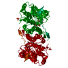
-Non-polymers , 6 types, 223 molecules 


| #2: Chemical | ChemComp-UDP /  Type: RNA linking / Mass: 404.161 Da / Num. of mol.: 1 / Source method: obtained synthetically / Formula: C9H14N2O12P2 / Comment: UDP*YM Type: RNA linking / Mass: 404.161 Da / Num. of mol.: 1 / Source method: obtained synthetically / Formula: C9H14N2O12P2 / Comment: UDP*YM | ||||
|---|---|---|---|---|---|
| #3: Chemical | ChemComp-MN /  Mass: 54.938 Da / Num. of mol.: 1 / Source method: obtained synthetically / Formula: Mn Mass: 54.938 Da / Num. of mol.: 1 / Source method: obtained synthetically / Formula: Mn | ||||
| #4: Chemical | ChemComp-3PG /  Mass: 186.057 Da / Num. of mol.: 1 / Source method: obtained synthetically / Formula: C3H7O7P Mass: 186.057 Da / Num. of mol.: 1 / Source method: obtained synthetically / Formula: C3H7O7P | ||||
| #5: Chemical |  Mass: 94.971 Da / Num. of mol.: 2 / Source method: obtained synthetically / Formula: PO4 Mass: 94.971 Da / Num. of mol.: 2 / Source method: obtained synthetically / Formula: PO4#6: Chemical | ChemComp-GOL / |  Mass: 92.094 Da / Num. of mol.: 1 / Source method: obtained synthetically / Formula: C3H8O3 Mass: 92.094 Da / Num. of mol.: 1 / Source method: obtained synthetically / Formula: C3H8O3#7: Water | ChemComp-HOH / | Mass: 18.015 Da / Num. of mol.: 217 / Source method: isolated from a natural source / Formula: H2O |
-Experimental details
-Experiment
| Experiment | Method: X-RAY DIFFRACTION / Number of used crystals: 1 |
|---|
- Sample preparation
| Crystal | Density Matthews: 4.37 Å3/Da / Density % sol: 71.84 % |
|---|---|
| Crystal grow | Temperature: 291 K / Method: vapor diffusion, sitting drop / pH: 7.5 Details: 5mM UDP, 5mM 3PG, 5mM MnCl2, Tris-HCL pH 7.5, VAPOR DIFFUSION, SITTING DROP, temperature 291K |
-Data collection
| Diffraction | Mean temperature: 100 K |
|---|---|
| Diffraction source | Source: SYNCHROTRON / Site: ESRF  / Beamline: ID23-2 / Wavelength: 0.8726 Å / Beamline: ID23-2 / Wavelength: 0.8726 Å |
| Detector | Type: MARMOSAIC 225 mm CCD / Detector: CCD / Date: Jul 8, 2011 |
| Radiation | Monochromator: Si 111 CHANNEL / Protocol: SINGLE WAVELENGTH / Monochromatic (M) / Laue (L): M / Scattering type: x-ray |
| Radiation wavelength | Wavelength: 0.8726 Å / Relative weight: 1 |
| Reflection | Resolution: 1.98→39.36 Å / Num. all: 43786 / Num. obs: 42409 / % possible obs: 96.9 % / Observed criterion σ(F): 0 / Observed criterion σ(I): -3 / Redundancy: 3.8 % / Biso Wilson estimate: 33.71 Å2 / Rmerge(I) obs: 0.062 / Net I/σ(I): 12.4 |
| Reflection shell | Resolution: 1.98→2.1 Å / Redundancy: 3.8 % / Rmerge(I) obs: 0.498 / Mean I/σ(I) obs: 2.02 / Num. unique all: 6950 / % possible all: 98.4 |
-Phasing
| Phasing | Method: molecular replacement |
|---|
- Processing
| Software |
| |||||||||||||||||||||||||||||||||||||||||||||||||||||||||||||||||||||||||||||||||||||||||||||||||||||||||
|---|---|---|---|---|---|---|---|---|---|---|---|---|---|---|---|---|---|---|---|---|---|---|---|---|---|---|---|---|---|---|---|---|---|---|---|---|---|---|---|---|---|---|---|---|---|---|---|---|---|---|---|---|---|---|---|---|---|---|---|---|---|---|---|---|---|---|---|---|---|---|---|---|---|---|---|---|---|---|---|---|---|---|---|---|---|---|---|---|---|---|---|---|---|---|---|---|---|---|---|---|---|---|---|---|---|---|
| Refinement | Method to determine structure: MOLECULAR REPLACEMENT / Resolution: 1.98→39.36 Å / Occupancy max: 1 / Occupancy min: 0.28 / FOM work R set: 0.853 / SU ML: 0.55 / σ(F): 2.01 / Phase error: 22.2 / Stereochemistry target values: ML
| |||||||||||||||||||||||||||||||||||||||||||||||||||||||||||||||||||||||||||||||||||||||||||||||||||||||||
| Solvent computation | Shrinkage radii: 1.24 Å / VDW probe radii: 1.4 Å / Solvent model: FLAT BULK SOLVENT MODEL / Bsol: 57.412 Å2 / ksol: 0.352 e/Å3 | |||||||||||||||||||||||||||||||||||||||||||||||||||||||||||||||||||||||||||||||||||||||||||||||||||||||||
| Displacement parameters | Biso max: 109.08 Å2 / Biso mean: 41.9334 Å2 / Biso min: 19.25 Å2
| |||||||||||||||||||||||||||||||||||||||||||||||||||||||||||||||||||||||||||||||||||||||||||||||||||||||||
| Refinement step | Cycle: LAST / Resolution: 1.98→39.36 Å
| |||||||||||||||||||||||||||||||||||||||||||||||||||||||||||||||||||||||||||||||||||||||||||||||||||||||||
| Refine LS restraints |
| |||||||||||||||||||||||||||||||||||||||||||||||||||||||||||||||||||||||||||||||||||||||||||||||||||||||||
| LS refinement shell | Refine-ID: X-RAY DIFFRACTION / Total num. of bins used: 14
|
