 Movie
Movie Controller

[English] 日本語
 Yorodumi
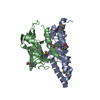
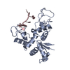
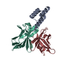
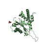
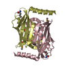
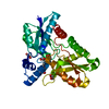
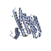
- PDB-4day: Crystal structure of the RMI core complex with MM2 peptide from FANCM -
+ Open data

- Basic information
| Entry | Database: PDB / ID: 4day | ||||||
|---|---|---|---|---|---|---|---|
| Title | Crystal structure of the RMI core complex with MM2 peptide from FANCM | ||||||
 Components Components |
| ||||||
 Keywords Keywords | protein binding/Hydrolase / OB fold / protein binding-Hydrolase complex | ||||||
| Function / homology |  Function and homology information Function and homology informationregulation of sister chromatid segregation / RecQ family helicase-topoisomerase III complex / double-strand break repair via synthesis-dependent strand annealing / FANCM-MHF complex / reduction of food intake in response to dietary excess / resolution of DNA recombination intermediates / Fanconi anaemia nuclear complex / maintenance of rDNA / resolution of meiotic recombination intermediates / four-way junction helicase activity ...regulation of sister chromatid segregation / RecQ family helicase-topoisomerase III complex / double-strand break repair via synthesis-dependent strand annealing / FANCM-MHF complex / reduction of food intake in response to dietary excess / resolution of DNA recombination intermediates / Fanconi anaemia nuclear complex / maintenance of rDNA / resolution of meiotic recombination intermediates / four-way junction helicase activity / nuclease activity / Impaired BRCA2 binding to PALB2 / positive regulation of protein monoubiquitination / HDR through Single Strand Annealing (SSA) / Homologous DNA Pairing and Strand Exchange / Defective homologous recombination repair (HRR) due to BRCA1 loss of function / Defective HDR through Homologous Recombination Repair (HRR) due to PALB2 loss of BRCA1 binding function / Defective HDR through Homologous Recombination Repair (HRR) due to PALB2 loss of BRCA2/RAD51/RAD51C binding function / Resolution of D-loop Structures through Synthesis-Dependent Strand Annealing (SDSA) / Resolution of D-loop Structures through Holliday Junction Intermediates / 3'-5' DNA helicase activity / Impaired BRCA2 binding to RAD51 / replication fork processing / Presynaptic phase of homologous DNA pairing and strand exchange / negative regulation of double-strand break repair via homologous recombination / response to glucose / interstrand cross-link repair / four-way junction DNA binding / Fanconi Anemia Pathway / PKR-mediated signaling / G2/M DNA damage checkpoint / double-strand break repair via homologous recombination / multicellular organism growth / HDR through Homologous Recombination (HRR) / glucose homeostasis / Processing of DNA double-strand break ends / Regulation of TP53 Activity through Phosphorylation / DNA replication / RNA helicase activity / nuclear speck / nuclear body / RNA helicase / nucleotide binding / DNA repair / chromatin binding / chromatin / ATP hydrolysis activity / DNA binding / nucleoplasm / ATP binding / nucleus / cytosol Similarity search - Function | ||||||
| Biological species |  Homo sapiens (human) Homo sapiens (human) | ||||||
| Method |  X-RAY DIFFRACTION / SYNCHROTRON / MOLECULAR REPLACEMENT / Resolution: 3.3 Å X-RAY DIFFRACTION / SYNCHROTRON / MOLECULAR REPLACEMENT / Resolution: 3.3 Å | ||||||
 Authors Authors | Hoadley, K.A. / Keck, J.L. | ||||||
 Citation Citation | Journal: Proc.Natl.Acad.Sci.USA / Year: 2012 Title: Defining the molecular interface that connects the Fanconi anemia protein FANCM to the Bloom syndrome dissolvasome. Authors: Hoadley, K.A. / Xue, Y. / Ling, C. / Takata, M. / Wang, W. / Keck, J.L. | ||||||
| History |
|
- Structure visualization
| Structure viewer | Molecule: MolmilJmol/JSmol |
|---|
- Downloads & links
-Download
| PDBx/mmCIF format | 4day.cif.gz | 121.3 KB | Display | PDBx/mmCIF format |
|---|---|---|---|---|
| PDB format | pdb4day.ent.gz | 94.3 KB | Display | PDB format |
| PDBx/mmJSON format | 4day.json.gz | Tree view | PDBx/mmJSON format | |
| Others |  Other downloads Other downloads |
-Validation report
| Arichive directory | https://data.pdbj.org/pub/pdb/validation_reports/da/4dayftp://data.pdbj.org/pub/pdb/validation_reports/da/4day | HTTPS FTP |
|---|
-Related structure data
| Related structure data | 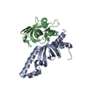 3mxnS S: Starting model for refinement |
|---|---|
| Similar structure data |


-Links
 PDBj
PDBj


- Assembly
| Deposited unit | 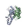
| ||||||||
|---|---|---|---|---|---|---|---|---|---|
| 1 |
| ||||||||
| Unit cell |
|
-Components
| #1: Protein | Mass: 17401.254 Da / Num. of mol.: 1 / Fragment: C-terminal OB domain (residues 473-625) Source method: isolated from a genetically manipulated source Source: (gene. exp.) Homo sapiens (human) / Gene: RMI1, C9orf76 / Production host:  |
|---|---|
| #2: Protein | Mass: 16168.581 Da / Num. of mol.: 1 Source method: isolated from a genetically manipulated source Source: (gene. exp.) Homo sapiens (human) / Gene: RMI2, C16orf75 / Production host: |
| #3: Protein/peptide | Mass: 4199.458 Da / Num. of mol.: 1 / Fragment: MM2 peptide (residues 1218-1251) Source method: isolated from a genetically manipulated source Source: (gene. exp.) Homo sapiens (human) / Gene: FANCM, KIAA1596 / Production host: |
-Experimental details
-Experiment
| Experiment | Method: X-RAY DIFFRACTION / Number of used crystals: 1 |
|---|
- Sample preparation
| Crystal | Density Matthews: 2.38 Å3/Da / Density % sol: 48.33 % |
|---|---|
| Crystal grow | Temperature: 298 K / Method: vapor diffusion, hanging drop / pH: 8.8 Details: 200 mM Na2SO4, 5% polyethylene glycol 3350, pH 8.8, VAPOR DIFFUSION, HANGING DROP, temperature 298K |
-Data collection
| Diffraction | Mean temperature: 100 K | |||||||||||||||||||||||||||||||||||||||||||||||||||||||||||||||||||||||||||||||||||||||||||||||||||||||||||||||||||||||||||||||||||||||||||||||||||
|---|---|---|---|---|---|---|---|---|---|---|---|---|---|---|---|---|---|---|---|---|---|---|---|---|---|---|---|---|---|---|---|---|---|---|---|---|---|---|---|---|---|---|---|---|---|---|---|---|---|---|---|---|---|---|---|---|---|---|---|---|---|---|---|---|---|---|---|---|---|---|---|---|---|---|---|---|---|---|---|---|---|---|---|---|---|---|---|---|---|---|---|---|---|---|---|---|---|---|---|---|---|---|---|---|---|---|---|---|---|---|---|---|---|---|---|---|---|---|---|---|---|---|---|---|---|---|---|---|---|---|---|---|---|---|---|---|---|---|---|---|---|---|---|---|---|---|---|---|
| Diffraction source | Source: SYNCHROTRON / Site: APS  / Beamline: 21-ID-F / Wavelength: 0.97856 Å / Beamline: 21-ID-F / Wavelength: 0.97856 Å | |||||||||||||||||||||||||||||||||||||||||||||||||||||||||||||||||||||||||||||||||||||||||||||||||||||||||||||||||||||||||||||||||||||||||||||||||||
| Detector | Type: MARMOSAIC 225 mm CCD / Detector: CCD / Date: Feb 13, 2011 | |||||||||||||||||||||||||||||||||||||||||||||||||||||||||||||||||||||||||||||||||||||||||||||||||||||||||||||||||||||||||||||||||||||||||||||||||||
| Radiation | Monochromator: Diamond [111] / Protocol: SINGLE WAVELENGTH / Monochromatic (M) / Laue (L): M / Scattering type: x-ray | |||||||||||||||||||||||||||||||||||||||||||||||||||||||||||||||||||||||||||||||||||||||||||||||||||||||||||||||||||||||||||||||||||||||||||||||||||
| Radiation wavelength | Wavelength: 0.97856 Å / Relative weight: 1 | |||||||||||||||||||||||||||||||||||||||||||||||||||||||||||||||||||||||||||||||||||||||||||||||||||||||||||||||||||||||||||||||||||||||||||||||||||
| Reflection | Resolution: 3.3→50 Å / Num. all: 5728 / Num. obs: 5711 / % possible obs: 99.7 % / Observed criterion σ(F): 0 / Observed criterion σ(I): 0 / Redundancy: 5.1 % / Rmerge(I) obs: 0.081 / Χ2: 1.163 / Net I/σ(I): 9.1 | |||||||||||||||||||||||||||||||||||||||||||||||||||||||||||||||||||||||||||||||||||||||||||||||||||||||||||||||||||||||||||||||||||||||||||||||||||
| Reflection shell |
|
- Processing
| Software |
| ||||||||||||||||||||||||||||||||||||||||||||||||||||||||||||||||||||||||||||||||||||||||||||||||||||
|---|---|---|---|---|---|---|---|---|---|---|---|---|---|---|---|---|---|---|---|---|---|---|---|---|---|---|---|---|---|---|---|---|---|---|---|---|---|---|---|---|---|---|---|---|---|---|---|---|---|---|---|---|---|---|---|---|---|---|---|---|---|---|---|---|---|---|---|---|---|---|---|---|---|---|---|---|---|---|---|---|---|---|---|---|---|---|---|---|---|---|---|---|---|---|---|---|---|---|---|---|---|
| Refinement | Method to determine structure: MOLECULAR REPLACEMENT Starting model: pdb entry 3mxn Resolution: 3.3→50 Å / Cor.coef. Fo:Fc: 0.931 / Cor.coef. Fo:Fc free: 0.82 / WRfactor Rfree: 0.3042 / WRfactor Rwork: 0.1997 / Occupancy max: 1 / Occupancy min: 1 / FOM work R set: 0.7471 / SU B: 75.396 / SU ML: 0.557 / SU R Cruickshank DPI: 0.4697 / SU Rfree: 0.6787 / Cross valid method: THROUGHOUT / σ(F): 0 / ESU R Free: 0.679 / Stereochemistry target values: MAXIMUM LIKELIHOOD Details: HYDROGENS HAVE BEEN ADDED IN THE RIDING POSITIONS U VALUES : RESIDUAL ONLY
| ||||||||||||||||||||||||||||||||||||||||||||||||||||||||||||||||||||||||||||||||||||||||||||||||||||
| Solvent computation | Ion probe radii: 0.8 Å / Shrinkage radii: 0.8 Å / VDW probe radii: 1.4 Å / Solvent model: MASK | ||||||||||||||||||||||||||||||||||||||||||||||||||||||||||||||||||||||||||||||||||||||||||||||||||||
| Displacement parameters | Biso max: 100.78 Å2 / Biso mean: 92.948 Å2 / Biso min: 23.86 Å2
| ||||||||||||||||||||||||||||||||||||||||||||||||||||||||||||||||||||||||||||||||||||||||||||||||||||
| Refinement step | Cycle: LAST / Resolution: 3.3→50 Å
| ||||||||||||||||||||||||||||||||||||||||||||||||||||||||||||||||||||||||||||||||||||||||||||||||||||
| Refine LS restraints |
| ||||||||||||||||||||||||||||||||||||||||||||||||||||||||||||||||||||||||||||||||||||||||||||||||||||
| LS refinement shell | Resolution: 3.286→3.371 Å / Total num. of bins used: 20
| ||||||||||||||||||||||||||||||||||||||||||||||||||||||||||||||||||||||||||||||||||||||||||||||||||||
| Refinement TLS params. | Method: refined / Refine-ID: X-RAY DIFFRACTION
| ||||||||||||||||||||||||||||||||||||||||||||||||||||||||||||||||||||||||||||||||||||||||||||||||||||
| Refinement TLS group |
|
