 Movie
Movie Controller

[English] 日本語
 Yorodumi
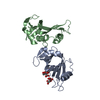
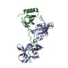
- PDB-4d01: Crystal Structure of the Extracellular Domain of the Human Alpha9... -
+ Open data

- Basic information
| Entry | Database: PDB / ID: 4d01 | ||||||
|---|---|---|---|---|---|---|---|
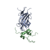
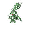
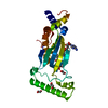
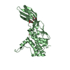
| Title | Crystal Structure of the Extracellular Domain of the Human Alpha9 Nicotinic Acetylcholine Receptor | ||||||
 Components Components | NEURONAL ACETYLCHOLINE RECEPTOR SUBUNIT ALPHA-9 | ||||||
 Keywords Keywords | SIGNALING PROTEIN / LIGAND BINDING DOMAIN / CYS-LOOP RECEPTOR | ||||||
| Function / homology |  Function and homology information Function and homology informationserotonin-gated monoatomic cation channel activity / Acetylcholine inhibits contraction of outer hair cells / cholinergic synapse / Highly calcium permeable postsynaptic nicotinic acetylcholine receptors / acetylcholine-gated channel complex / detection of mechanical stimulus involved in sensory perception of sound / response to auditory stimulus / acetylcholine-gated monoatomic cation-selective channel activity / transmembrane transporter complex / acetylcholine receptor signaling pathway ...serotonin-gated monoatomic cation channel activity / Acetylcholine inhibits contraction of outer hair cells / cholinergic synapse / Highly calcium permeable postsynaptic nicotinic acetylcholine receptors / acetylcholine-gated channel complex / detection of mechanical stimulus involved in sensory perception of sound / response to auditory stimulus / acetylcholine-gated monoatomic cation-selective channel activity / transmembrane transporter complex / acetylcholine receptor signaling pathway / excitatory extracellular ligand-gated monoatomic ion channel activity / inner ear morphogenesis / postsynaptic specialization membrane / membrane depolarization / transmitter-gated monoatomic ion channel activity involved in regulation of postsynaptic membrane potential / regulation of membrane potential / negative regulation of ERK1 and ERK2 cascade / calcium channel activity / positive regulation of cytosolic calcium ion concentration / monoatomic ion transmembrane transport / chemical synaptic transmission / neuron projection / synapse / plasma membrane Similarity search - Function | ||||||
| Biological species |  HOMO SAPIENS (human) HOMO SAPIENS (human) | ||||||
| Method |  X-RAY DIFFRACTION / SYNCHROTRON / MOLECULAR REPLACEMENT / Resolution: 1.795 Å X-RAY DIFFRACTION / SYNCHROTRON / MOLECULAR REPLACEMENT / Resolution: 1.795 Å | ||||||
 Authors Authors | Giastas, P. / Zouridakis, M. / Zarkadas, E. / Tzartos, S.J. | ||||||
 Citation Citation | Journal: Nat.Struct.Mol.Biol. / Year: 2014 Title: Crystal Structures of Free and Antagonist-Bound States of Human Alpha9 Nicotinic Receptor Extracellular Domain Authors: Zouridakis, M. / Giastas, P. / Zarkadas, E. / Chroni-Tzartou, D. / Bregestovski, P. / Tzartos, S.J. | ||||||
| History |
|
- Structure visualization
| Structure viewer | Molecule: MolmilJmol/JSmol |
|---|
- Downloads & links
-Download
| PDBx/mmCIF format | 4d01.cif.gz | 107.9 KB | Display | PDBx/mmCIF format |
|---|---|---|---|---|
| PDB format | pdb4d01.ent.gz | 84.4 KB | Display | PDB format |
| PDBx/mmJSON format | 4d01.json.gz | Tree view | PDBx/mmJSON format | |
| Others |  Other downloads Other downloads |
-Validation report
| Arichive directory | https://data.pdbj.org/pub/pdb/validation_reports/d0/4d01ftp://data.pdbj.org/pub/pdb/validation_reports/d0/4d01 | HTTPS FTP |
|---|
-Related structure data
| Related structure data |  4uxuC  4uy2C  2qc1S C: citing same article ( S: Starting model for refinement |
|---|---|
| Similar structure data |


-Links
 PDBj
PDBj

- Assembly
| Deposited unit | 
| ||||||||
|---|---|---|---|---|---|---|---|---|---|
| 1 |
| ||||||||
| Unit cell |
| ||||||||
| Components on special symmetry positions |
|
-Components
| #1: Protein | Mass: 25307.029 Da / Num. of mol.: 1 / Fragment: EXTRACELLULAR DOMAIN, RESIDUES 26-237 Source method: isolated from a genetically manipulated source Source: (gene. exp.) HOMO SAPIENS (human) / Plasmid: PPICZAA / Production host:  KOMAGATAELLA PASTORIS (fungus) / Strain (production host): X33 / References: UniProt: Q9UGM1 KOMAGATAELLA PASTORIS (fungus) / Strain (production host): X33 / References: UniProt: Q9UGM1 | ||||||||
|---|---|---|---|---|---|---|---|---|---|
| #2: Sugar |   Type: D-saccharide, beta linking / Mass: 221.208 Da / Num. of mol.: 2 Type: D-saccharide, beta linking / Mass: 221.208 Da / Num. of mol.: 2Source method: isolated from a genetically manipulated source Formula: C8H15NO6 #3: Chemical | ChemComp-EDO /   Mass: 62.068 Da / Num. of mol.: 4 / Source method: obtained synthetically / Formula: C2H6O2 Mass: 62.068 Da / Num. of mol.: 4 / Source method: obtained synthetically / Formula: C2H6O2#4: Water | ChemComp-HOH / |  Mass: 18.015 Da / Num. of mol.: 206 / Source method: isolated from a natural source / Formula: H2O Mass: 18.015 Da / Num. of mol.: 206 / Source method: isolated from a natural source / Formula: H2OHas protein modification | Y | Sequence details | EXTRACELLU | |
-Experimental details
-Experiment
| Experiment | Method: X-RAY DIFFRACTION / Number of used crystals: 1 |
|---|
- Sample preparation
| Crystal | Density Matthews: 2.4 Å3/Da / Density % sol: 51 % / Description: NONE |
|---|---|
| Crystal grow | pH: 7.5 / Details: 100 MM SPG BUFFER PH 6.5, 25% PEG 3350 |
-Data collection
| Diffraction | Mean temperature: 100 K |
|---|---|
| Diffraction source | Source: SYNCHROTRON / Site: SLS  / Beamline: X06DA / Wavelength: 1 / Beamline: X06DA / Wavelength: 1 |
| Detector | Type: DECTRIS PILATUS 2M / Detector: PIXEL / Date: Jan 10, 2013 |
| Radiation | Protocol: SINGLE WAVELENGTH / Monochromatic (M) / Laue (L): M / Scattering type: x-ray |
| Radiation wavelength | Wavelength: 1 Å / Relative weight: 1 |
| Reflection | Resolution: 1.79→42 Å / Num. obs: 24313 / % possible obs: 99.3 % / Observed criterion σ(I): 1.4 / Redundancy: 12.5 % / Biso Wilson estimate: 32.7 Å2 / Rmerge(I) obs: 0.08 / Net I/σ(I): 15.9 |
| Reflection shell | Resolution: 1.79→42 Å / Redundancy: 11.4 % / Rmerge(I) obs: 1.4 / Mean I/σ(I) obs: 1.2 / % possible all: 95.4 |
- Processing
| Software |
| |||||||||||||||||||||||||||||||||||||||||||||||||||||||||||||||||||||||||||||||||||||||||||||||||||||||||||||||||||||||||||||
|---|---|---|---|---|---|---|---|---|---|---|---|---|---|---|---|---|---|---|---|---|---|---|---|---|---|---|---|---|---|---|---|---|---|---|---|---|---|---|---|---|---|---|---|---|---|---|---|---|---|---|---|---|---|---|---|---|---|---|---|---|---|---|---|---|---|---|---|---|---|---|---|---|---|---|---|---|---|---|---|---|---|---|---|---|---|---|---|---|---|---|---|---|---|---|---|---|---|---|---|---|---|---|---|---|---|---|---|---|---|---|---|---|---|---|---|---|---|---|---|---|---|---|---|---|---|---|
| Refinement | Method to determine structure: MOLECULAR REPLACEMENT Starting model: PDB ENTRY 2QC1 Resolution: 1.795→42.126 Å / SU ML: 0.24 / σ(F): 1.33 / Phase error: 28.07 / Stereochemistry target values: ML Details: RESIDUES OF THE REGION 100-104 WAS EITHER LEFT UNMODELED OR WAS MODELED ONLY THEIR MAIN CHAIN ATOMS
| |||||||||||||||||||||||||||||||||||||||||||||||||||||||||||||||||||||||||||||||||||||||||||||||||||||||||||||||||||||||||||||
| Solvent computation | Shrinkage radii: 0.9 Å / VDW probe radii: 1.11 Å / Solvent model: FLAT BULK SOLVENT MODEL | |||||||||||||||||||||||||||||||||||||||||||||||||||||||||||||||||||||||||||||||||||||||||||||||||||||||||||||||||||||||||||||
| Displacement parameters | Biso mean: 48 Å2 | |||||||||||||||||||||||||||||||||||||||||||||||||||||||||||||||||||||||||||||||||||||||||||||||||||||||||||||||||||||||||||||
| Refinement step | Cycle: LAST / Resolution: 1.795→42.126 Å
| |||||||||||||||||||||||||||||||||||||||||||||||||||||||||||||||||||||||||||||||||||||||||||||||||||||||||||||||||||||||||||||
| Refine LS restraints |
| |||||||||||||||||||||||||||||||||||||||||||||||||||||||||||||||||||||||||||||||||||||||||||||||||||||||||||||||||||||||||||||
| LS refinement shell |
| |||||||||||||||||||||||||||||||||||||||||||||||||||||||||||||||||||||||||||||||||||||||||||||||||||||||||||||||||||||||||||||
| Refinement TLS params. | Method: refined / Refine-ID: X-RAY DIFFRACTION
| |||||||||||||||||||||||||||||||||||||||||||||||||||||||||||||||||||||||||||||||||||||||||||||||||||||||||||||||||||||||||||||
| Refinement TLS group |
|
