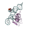
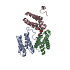
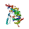
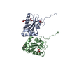
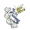
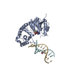
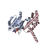
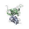
Entry Database : PDB / ID : 2qc1Title Crystal structure of the extracellular domain of the nicotinic acetylcholine receptor 1 subunit bound to alpha-bungarotoxin at 1.9 A resolution Acetylcholine receptor subunit alpha Alpha-bungarotoxin Keywords / / / / / Function / homology Function Domain/homology Component
/ / / / / / / / / / / / / / / / / / / / / / / / / / / / / / / / / / / / / / / / / / / / / / / / / / / / / / / / / / / / / / / / / / / / / / / / / / / / / Biological species Mus musculus (house mouse)Bungarus multicinctus (many-banded krait)Method / / / Resolution : 1.94 Å Authors Dellisanti, C.D. / Yao, Y. / Stroud, J.C. / Wang, Z. / Chen, L. Journal : Nat.Neurosci. / Year : 2007Title : Crystal structure of the extracellular domain of nAChR alpha1 bound to alpha-bungarotoxin at 1.94 A resolution.Authors : Dellisanti, C.D. / Yao, Y. / Stroud, J.C. / Wang, Z.Z. / Chen, L. History Deposition Jun 18, 2007 Deposition site / Processing site Revision 1.0 Aug 7, 2007 Provider / Type Revision 1.1 Jul 13, 2011 Group / Version format complianceRevision 2.0 Jul 29, 2020 Group Advisory / Atomic model ... Advisory / Atomic model / Data collection / Database references / Derived calculations / Structure summary Category atom_site / chem_comp ... atom_site / chem_comp / database_PDB_caveat / entity / pdbx_branch_scheme / pdbx_chem_comp_identifier / pdbx_entity_branch / pdbx_entity_branch_descriptor / pdbx_entity_branch_link / pdbx_entity_branch_list / pdbx_entity_nonpoly / pdbx_nonpoly_scheme / pdbx_struct_assembly_gen / pdbx_validate_chiral / struct_asym / struct_conn / struct_ref_seq_dif / struct_site / struct_site_gen Item _atom_site.auth_asym_id / _atom_site.auth_seq_id ... _atom_site.auth_asym_id / _atom_site.auth_seq_id / _atom_site.label_asym_id / _atom_site.label_entity_id / _chem_comp.name / _chem_comp.type / _pdbx_struct_assembly_gen.asym_id_list / _pdbx_validate_chiral.auth_asym_id / _pdbx_validate_chiral.auth_seq_id / _struct_conn.pdbx_dist_value / _struct_conn.pdbx_leaving_atom_flag / _struct_conn.pdbx_role / _struct_conn.ptnr1_auth_asym_id / _struct_conn.ptnr1_auth_seq_id / _struct_conn.ptnr1_label_asym_id / _struct_conn.ptnr1_label_atom_id / _struct_conn.ptnr2_auth_asym_id / _struct_conn.ptnr2_auth_seq_id / _struct_conn.ptnr2_label_asym_id / _struct_conn.ptnr2_label_seq_id / _struct_ref_seq_dif.details Description / Provider / Type Revision 2.1 Oct 20, 2021 Group / Structure summary / Category / database_2 / struct_ref_seq_difItem _chem_comp.pdbx_synonyms / _database_2.pdbx_DOI ... _chem_comp.pdbx_synonyms / _database_2.pdbx_DOI / _database_2.pdbx_database_accession / _struct_ref_seq_dif.details Revision 2.2 Aug 30, 2023 Group / Refinement descriptionCategory / chem_comp_bond / pdbx_initial_refinement_modelRevision 2.3 Oct 30, 2024 Group / Category / pdbx_modification_feature
Show all Show less
 Movie
Movie Controller

 Yorodumi
 Open data
 Basic information
 Components
 Keywords
 Function and homology information
 Bungarus multicinctus (many-banded krait)
 X-RAY DIFFRACTION /
  Authors
 Citation
 Structure visualization
 Downloads & links
 Other downloads
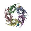

 PDBj
PDBj


 Assembly
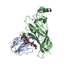

 Mass: 18.015 Da / Num. of mol.: 187 / Source method: isolated from a natural source / Formula: H2O
 Sample preparation
 / Beamline: 8.2.1 / Wavelength: 1 Å
 Processing