 Movie
Movie Controller


+ Open data

- Basic information
| Entry | Database: PDB / ID: 4czm | |||||||||
|---|---|---|---|---|---|---|---|---|---|---|
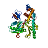
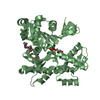
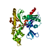
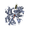
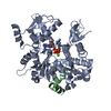
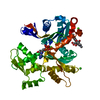
| Title | C. crescentus MreB, monomeric, AMPPNP | |||||||||
 Components Components | ROD SHAPE-DETERMINING PROTEIN MREB | |||||||||
 Keywords Keywords | STRUCTURAL PROTEIN / BACTERIAL ACTIN / BACTERIAL CYTOSKELETON | |||||||||
| Function / homology |  Function and homology information Function and homology information | |||||||||
| Biological species |  CAULOBACTER VIBRIOIDES (bacteria) CAULOBACTER VIBRIOIDES (bacteria) | |||||||||
| Method |  X-RAY DIFFRACTION / SYNCHROTRON / MOLECULAR REPLACEMENT / Resolution: 2.2 Å X-RAY DIFFRACTION / SYNCHROTRON / MOLECULAR REPLACEMENT / Resolution: 2.2 Å | |||||||||
 Authors Authors | Lowe, J. / van den Ent, F. | |||||||||
 Citation Citation | Journal: Elife / Year: 2014 Title: Bacterial Actin Mreb Forms Antiparallel Double Filaments. Authors: Van Den Ent, F. / Izore, T. / Bharat, T.A. / Johnson, C.M. / Lowe, J. | |||||||||
| History |
|
- Structure visualization
| Structure viewer | Molecule: MolmilJmol/JSmol |
|---|
- Downloads & links
-Download
| PDBx/mmCIF format | 4czm.cif.gz | 251.9 KB | Display | PDBx/mmCIF format |
|---|---|---|---|---|
| PDB format | pdb4czm.ent.gz | 205 KB | Display | PDB format |
| PDBx/mmJSON format | 4czm.json.gz | Tree view | PDBx/mmJSON format | |
| Others |  Other downloads Other downloads |
-Validation report
| Arichive directory | https://data.pdbj.org/pub/pdb/validation_reports/cz/4czmftp://data.pdbj.org/pub/pdb/validation_reports/cz/4czm | HTTPS FTP |
|---|
-Related structure data
| Related structure data | 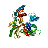 4czeC  4czfC 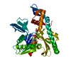 4czgC 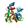 4czhC 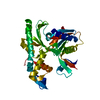 4cziC 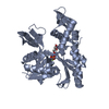 4czjC 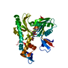 4czkC 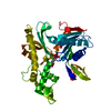 4czlC 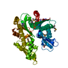 1jceS C: citing same article ( S: Starting model for refinement |
|---|---|
| Similar structure data |


-Links
 PDBj
PDBj


- Assembly
| Deposited unit | 
| ||||||||
|---|---|---|---|---|---|---|---|---|---|
| 1 | 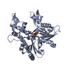
| ||||||||
| 2 | 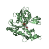
| ||||||||
| Unit cell |
| ||||||||
| Components on special symmetry positions |
|
-Components
| #1: Protein | Mass: 36893.504 Da / Num. of mol.: 2 / Fragment: RESIDUES 9-347 / Mutation: YES Source method: isolated from a genetically manipulated source Source: (gene. exp.) CAULOBACTER VIBRIOIDES (bacteria) / Plasmid: PHIS17 / Production host: #2: Chemical |   Mass: 24.305 Da / Num. of mol.: 2 / Source method: obtained synthetically / Formula: Mg Mass: 24.305 Da / Num. of mol.: 2 / Source method: obtained synthetically / Formula: Mg#3: Chemical |   Mass: 506.196 Da / Num. of mol.: 2 / Source method: obtained synthetically / Formula: C10H17N6O12P3 / Comment: AMP-PNP, energy-carrying molecule analogue*YM Mass: 506.196 Da / Num. of mol.: 2 / Source method: obtained synthetically / Formula: C10H17N6O12P3 / Comment: AMP-PNP, energy-carrying molecule analogue*YM#4: Water | ChemComp-HOH / |  Mass: 18.015 Da / Num. of mol.: 43 / Source method: isolated from a natural source / Formula: H2O Mass: 18.015 Da / Num. of mol.: 43 / Source method: isolated from a natural source / Formula: H2O |
|---|
-Experimental details
-Experiment
| Experiment | Method: X-RAY DIFFRACTION / Number of used crystals: 1 |
|---|
- Sample preparation
| Crystal | Density Matthews: 2.5 Å3/Da / Density % sol: 50.71 % / Description: NONE |
|---|
-Data collection
| Diffraction | Mean temperature: 100 K |
|---|---|
| Diffraction source | Source: SYNCHROTRON / Site: Diamond  / Beamline: I02 / Wavelength: 0.97957 / Beamline: I02 / Wavelength: 0.97957 |
| Detector | Type: DECTRIS PILATUS / Detector: PIXEL |
| Radiation | Protocol: SINGLE WAVELENGTH / Monochromatic (M) / Laue (L): M / Scattering type: x-ray |
| Radiation wavelength | Wavelength: 0.97957 Å / Relative weight: 1 |
| Reflection | Resolution: 2.2→50 Å / Num. obs: 39419 / % possible obs: 100 % / Observed criterion σ(I): 0 / Redundancy: 12.9 % / Biso Wilson estimate: 60.4 Å2 / Rmerge(I) obs: 0.06 / Net I/σ(I): 19.5 |
| Reflection shell | Resolution: 2.2→2.32 Å / Redundancy: 12.9 % / Rmerge(I) obs: 1.5 / Mean I/σ(I) obs: 1.8 / % possible all: 100 |
- Processing
| Software |
| |||||||||||||||||||||||||||||||||||||||||||||||||||||||||||||||||||||||||||||||||||||||||||||||||||||||||||||||||||||||||||||||||||||||||||||||||||||||||||||||||||||||||||||||||||||||||||||
|---|---|---|---|---|---|---|---|---|---|---|---|---|---|---|---|---|---|---|---|---|---|---|---|---|---|---|---|---|---|---|---|---|---|---|---|---|---|---|---|---|---|---|---|---|---|---|---|---|---|---|---|---|---|---|---|---|---|---|---|---|---|---|---|---|---|---|---|---|---|---|---|---|---|---|---|---|---|---|---|---|---|---|---|---|---|---|---|---|---|---|---|---|---|---|---|---|---|---|---|---|---|---|---|---|---|---|---|---|---|---|---|---|---|---|---|---|---|---|---|---|---|---|---|---|---|---|---|---|---|---|---|---|---|---|---|---|---|---|---|---|---|---|---|---|---|---|---|---|---|---|---|---|---|---|---|---|---|---|---|---|---|---|---|---|---|---|---|---|---|---|---|---|---|---|---|---|---|---|---|---|---|---|---|---|---|---|---|---|---|---|
| Refinement | Method to determine structure: MOLECULAR REPLACEMENT Starting model: PDB ENTRY 1JCE Resolution: 2.2→41.138 Å / SU ML: 0.39 / σ(F): 1.21 / Phase error: 30.79 / Stereochemistry target values: ML
| |||||||||||||||||||||||||||||||||||||||||||||||||||||||||||||||||||||||||||||||||||||||||||||||||||||||||||||||||||||||||||||||||||||||||||||||||||||||||||||||||||||||||||||||||||||||||||||
| Solvent computation | Shrinkage radii: 0.9 Å / VDW probe radii: 1.11 Å / Solvent model: FLAT BULK SOLVENT MODEL | |||||||||||||||||||||||||||||||||||||||||||||||||||||||||||||||||||||||||||||||||||||||||||||||||||||||||||||||||||||||||||||||||||||||||||||||||||||||||||||||||||||||||||||||||||||||||||||
| Refinement step | Cycle: LAST / Resolution: 2.2→41.138 Å
| |||||||||||||||||||||||||||||||||||||||||||||||||||||||||||||||||||||||||||||||||||||||||||||||||||||||||||||||||||||||||||||||||||||||||||||||||||||||||||||||||||||||||||||||||||||||||||||
| Refine LS restraints |
| |||||||||||||||||||||||||||||||||||||||||||||||||||||||||||||||||||||||||||||||||||||||||||||||||||||||||||||||||||||||||||||||||||||||||||||||||||||||||||||||||||||||||||||||||||||||||||||
| LS refinement shell |
|
