 Movie
Movie Controller

[English] 日本語
 Yorodumi
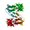
- PDB-4cxa: Crystal structure of the human CDK12-cyclin K complex bound to AMPPNP -
+ Open data

- Basic information
| Entry | Database: PDB / ID: 4cxa | ||||||
|---|---|---|---|---|---|---|---|
| Title | Crystal structure of the human CDK12-cyclin K complex bound to AMPPNP | ||||||
 Components Components |
| ||||||
 Keywords Keywords | TRANSFERASE / KINASE | ||||||
| Function / homology |  Function and homology information Function and homology informationcyclin K-CDK12 complex / cyclin K-CDK13 complex / nuclear cyclin-dependent protein kinase holoenzyme complex / regulation of MAP kinase activity / cyclin/CDK positive transcription elongation factor complex / host-mediated suppression of viral genome replication / negative regulation of intracellular estrogen receptor signaling pathway / negative regulation of stem cell differentiation / regulation of cyclin-dependent protein serine/threonine kinase activity / cyclin-dependent protein serine/threonine kinase activator activity ...cyclin K-CDK12 complex / cyclin K-CDK13 complex / nuclear cyclin-dependent protein kinase holoenzyme complex / regulation of MAP kinase activity / cyclin/CDK positive transcription elongation factor complex / host-mediated suppression of viral genome replication / negative regulation of intracellular estrogen receptor signaling pathway / negative regulation of stem cell differentiation / regulation of cyclin-dependent protein serine/threonine kinase activity / cyclin-dependent protein serine/threonine kinase activator activity / positive regulation of DNA-templated transcription, elongation / [RNA-polymerase]-subunit kinase / RNA polymerase II transcribes snRNA genes / cellular response to estrogen stimulus / HIV elongation arrest and recovery / Pausing and recovery of HIV elongation / regulation of signal transduction / cyclin-dependent kinase / cyclin-dependent protein serine/threonine kinase activity / Formation of HIV elongation complex in the absence of HIV Tat / RNA Polymerase II Transcription Elongation / Formation of RNA Pol II elongation complex / negative regulation of MAPK cascade / RNA polymerase II CTD heptapeptide repeat kinase activity / RNA Polymerase II Pre-transcription Events / RNA splicing / cyclin binding / TP53 Regulates Transcription of DNA Repair Genes / positive regulation of transcription elongation by RNA polymerase II / SMAD2/SMAD3:SMAD4 heterotrimer regulates transcription / mRNA processing / transcription by RNA polymerase II / protein kinase activity / nuclear speck / protein serine kinase activity / cell division / DNA damage response / protein kinase binding / positive regulation of transcription by RNA polymerase II / nucleoplasm / ATP binding / nucleus Similarity search - Function | ||||||
| Biological species |  HOMO SAPIENS (human) HOMO SAPIENS (human) | ||||||
| Method |  X-RAY DIFFRACTION / SYNCHROTRON / MOLECULAR REPLACEMENT / Resolution: 3.15 Å X-RAY DIFFRACTION / SYNCHROTRON / MOLECULAR REPLACEMENT / Resolution: 3.15 Å | ||||||
 Authors Authors | Dixon Clarke, S.E. / Elkins, J.M. / Pike, A.C.W. / Nowak, R. / Goubin, S. / Mahajan, R.P. / Kopec, J. / Froese, S. / Tallant, C. / Carpenter, E.P. ...Dixon Clarke, S.E. / Elkins, J.M. / Pike, A.C.W. / Nowak, R. / Goubin, S. / Mahajan, R.P. / Kopec, J. / Froese, S. / Tallant, C. / Carpenter, E.P. / Mackenzie, A. / Faust, B. / Burgess-Brown, N. / von Delft, F. / Arrowsmith, C. / Edwards, A.M. / Bountra, C. / Bullock, A. | ||||||
 Citation Citation | Journal: Sci.Rep. / Year: 2015 Title: Structures of the Cdk12/Cyck Complex with AMP-Pnp Reveal a Flexible C-Terminal Kinase Extension Important for ATP Binding. Authors: Dixon-Clarke, S.E. / Elkins, J.M. / Cheng, S.G. / Morin, G.B. / Bullock, A.N. | ||||||
| History |
|
- Structure visualization
| Structure viewer | Molecule: MolmilJmol/JSmol |
|---|
- Downloads & links
-Download
| PDBx/mmCIF format | 4cxa.cif.gz | 232.5 KB | Display | PDBx/mmCIF format |
|---|---|---|---|---|
| PDB format | pdb4cxa.ent.gz | 179.1 KB | Display | PDB format |
| PDBx/mmJSON format | 4cxa.json.gz | Tree view | PDBx/mmJSON format | |
| Others |  Other downloads Other downloads |
-Validation report
| Arichive directory | https://data.pdbj.org/pub/pdb/validation_reports/cx/4cxaftp://data.pdbj.org/pub/pdb/validation_reports/cx/4cxa | HTTPS FTP |
|---|
-Related structure data
| Related structure data |  4un0C  4cjy S: Starting model for refinement C: citing same article ( |
|---|---|
| Similar structure data |


-Links
 PDBj
PDBj


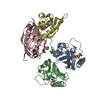
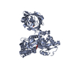
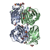
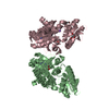
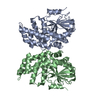
- Assembly
| Deposited unit | 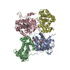
| ||||||||||||
|---|---|---|---|---|---|---|---|---|---|---|---|---|---|
| 1 | 
| ||||||||||||
| 2 | 
| ||||||||||||
| Unit cell |
| ||||||||||||
| Noncrystallographic symmetry (NCS) | NCS oper:
|
-Components
| #1: Protein | Mass: 39719.922 Da / Num. of mol.: 2 / Fragment: KINASE DOMAIN, RESIDUES 715-1052 Source method: isolated from a genetically manipulated source Source: (gene. exp.) HOMO SAPIENS (human) / Plasmid: PFB-LIC-BSE / Cell line (production host): SF9 / Production host:   SPODOPTERA FRUGIPERDA (fall armyworm) / References: UniProt: Q9NYV4, cyclin-dependent kinase SPODOPTERA FRUGIPERDA (fall armyworm) / References: UniProt: Q9NYV4, cyclin-dependent kinase#2: Protein | Mass: 30443.148 Da / Num. of mol.: 2 / Fragment: CYCLIN K, RESIDUES 11-267 Source method: isolated from a genetically manipulated source Source: (gene. exp.) HOMO SAPIENS (human) / Plasmid: PFB-LIC-BSE / Cell line (production host): SF9 / Production host: SPODOPTERA FRUGIPERDA (fall armyworm) / References: UniProt: O75909#3: Chemical | ChemComp-ANP / |   Mass: 506.196 Da / Num. of mol.: 1 / Source method: obtained synthetically / Formula: C10H17N6O12P3 / Comment: AMP-PNP, energy-carrying molecule analogue*YM Mass: 506.196 Da / Num. of mol.: 1 / Source method: obtained synthetically / Formula: C10H17N6O12P3 / Comment: AMP-PNP, energy-carrying molecule analogue*YMHas protein modification | Y | |
|---|
-Experimental details
-Experiment
| Experiment | Method: X-RAY DIFFRACTION / Number of used crystals: 1 |
|---|
- Sample preparation
| Crystal | Density Matthews: 2.22 Å3/Da / Density % sol: 48.72 % / Description: NONE |
|---|---|
| Crystal grow | pH: 7 / Details: 0.15M DL-MALIC ACID, 20% PEG3350, pH 7 |
-Data collection
| Diffraction | Mean temperature: 100 K |
|---|---|
| Diffraction source | Source: SYNCHROTRON / Site: Diamond  / Beamline: I02 / Wavelength: 0.97868 / Beamline: I02 / Wavelength: 0.97868 |
| Detector | Type: DECTRIS PILATUS 6M / Detector: PIXEL / Date: Feb 7, 2014 |
| Radiation | Protocol: SINGLE WAVELENGTH / Monochromatic (M) / Laue (L): M / Scattering type: x-ray |
| Radiation wavelength | Wavelength: 0.97868 Å / Relative weight: 1 |
| Reflection | Resolution: 3.15→30.53 Å / Num. obs: 22908 / % possible obs: 99.8 % / Observed criterion σ(I): 0 / Redundancy: 5 % / Rmerge(I) obs: 0.19 / Net I/σ(I): 6.4 |
| Reflection shell | Resolution: 3.15→3.37 Å / Redundancy: 4.9 % / Rmerge(I) obs: 0.98 / Mean I/σ(I) obs: 1.5 / % possible all: 99.9 |
- Processing
| Software |
| |||||||||||||||||||||||||||||||||||||||||||||||||||||||||||||||
|---|---|---|---|---|---|---|---|---|---|---|---|---|---|---|---|---|---|---|---|---|---|---|---|---|---|---|---|---|---|---|---|---|---|---|---|---|---|---|---|---|---|---|---|---|---|---|---|---|---|---|---|---|---|---|---|---|---|---|---|---|---|---|---|---|
| Refinement | Method to determine structure: MOLECULAR REPLACEMENT Starting model: PDB ENTRY 4CJY 4cjy Resolution: 3.15→30.529 Å / SU ML: 0.47 / σ(F): 1.35 / Phase error: 32.91 / Stereochemistry target values: ML
| |||||||||||||||||||||||||||||||||||||||||||||||||||||||||||||||
| Solvent computation | Shrinkage radii: 0.9 Å / VDW probe radii: 1.11 Å / Solvent model: FLAT BULK SOLVENT MODEL | |||||||||||||||||||||||||||||||||||||||||||||||||||||||||||||||
| Refinement step | Cycle: LAST / Resolution: 3.15→30.529 Å
| |||||||||||||||||||||||||||||||||||||||||||||||||||||||||||||||
| Refine LS restraints |
| |||||||||||||||||||||||||||||||||||||||||||||||||||||||||||||||
| LS refinement shell |
|
