 Movie
Movie Controller

[English] 日本語
 Yorodumi
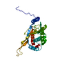
- PDB-3d2y: Complex of the N-acetylmuramyl-L-alanine amidase AmiD from E.coli... -
+ Open data

- Basic information
| Entry | Database: PDB / ID: 3d2y | ||||||
|---|---|---|---|---|---|---|---|
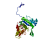
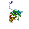
| Title | Complex of the N-acetylmuramyl-L-alanine amidase AmiD from E.coli with the substrate anhydro-N-acetylmuramic acid-L-Ala-D-gamma-Glu-L-Lys | ||||||
 Components Components |
| ||||||
 Keywords Keywords | HYDROLASE / ZINC AMIDASE / PGRP / Peptidoglycan Recognizing Protein / AmpD / N-ACETYLMURAMYL-L-ALANINE AMIDASE / Cell wall biogenesis/degradation / Lipoprotein / Membrane / Metal-binding / Outer membrane / Palmitate | ||||||
| Function / homology |  Function and homology information Function and homology informationN-acetyl-anhydromuramoyl-L-alanine amidase activity / N-acetylmuramoyl-L-alanine amidase / N-acetylmuramoyl-L-alanine amidase activity / peptidoglycan turnover / outer membrane / peptidoglycan catabolic process / cell outer membrane / cell wall organization / zinc ion binding Similarity search - Function | ||||||
| Biological species |  | ||||||
| Method |  X-RAY DIFFRACTION / SYNCHROTRON / MOLECULAR REPLACEMENT / Resolution: 1.75 Å X-RAY DIFFRACTION / SYNCHROTRON / MOLECULAR REPLACEMENT / Resolution: 1.75 Å | ||||||
 Authors Authors | Kerff, F. / Petrella, S. / Herman, R. / Sauvage, E. / Mercier, F. / Luxen, A. / Frere, J.M. / Joris, B. / Charlier, P. | ||||||
 Citation Citation | Journal: J.Mol.Biol. / Year: 2010 Title: Specific Structural Features of the N-Acetylmuramoyl-l-Alanine Amidase AmiD from Escherichia coli and Mechanistic Implications for Enzymes of This Family. Authors: Kerff, F. / Petrella, S. / Mercier, F. / Sauvage, E. / Herman, R. / Pennartz, A. / Zervosen, A. / Luxen, A. / Frere, J.M. / Joris, B. / Charlier, P. | ||||||
| History |
|
- Structure visualization
| Structure viewer | Molecule: MolmilJmol/JSmol |
|---|
- Downloads & links
-Download
| PDBx/mmCIF format | 3d2y.cif.gz | 76.6 KB | Display | PDBx/mmCIF format |
|---|---|---|---|---|
| PDB format | pdb3d2y.ent.gz | 57.1 KB | Display | PDB format |
| PDBx/mmJSON format | 3d2y.json.gz | Tree view | PDBx/mmJSON format | |
| Others |  Other downloads Other downloads |
-Validation report
| Arichive directory | https://data.pdbj.org/pub/pdb/validation_reports/d2/3d2yftp://data.pdbj.org/pub/pdb/validation_reports/d2/3d2y | HTTPS FTP |
|---|
-Related structure data
| Related structure data |  2bh7C  2wkxC  3d2zC  2bgx C: citing same article ( S: Starting model for refinement |
|---|---|
| Similar structure data |


-Links
 PDBj
PDBj


- Assembly
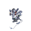
| Deposited unit | 
| ||||||||
|---|---|---|---|---|---|---|---|---|---|
| 1 |
| ||||||||
| 2 | 
| ||||||||
| Unit cell |
| ||||||||
| Components on special symmetry positions |
| ||||||||
| Details | The biological assembly is a monomer but the protein is found as a dimer in the crystal with a swapping of the N-terminus |
-Components
| #1: Protein | Mass: 29376.240 Da / Num. of mol.: 1 Source method: isolated from a genetically manipulated source Source: (gene. exp.) References: UniProt: P75820, N-acetylmuramoyl-L-alanine amidase | ||
|---|---|---|---|
| #2: Protein/peptide | Mass: 604.626 Da / Num. of mol.: 1 / Source method: obtained synthetically / Details: The peptide was chemically synthesized | ||
| #3: Chemical |   Mass: 92.094 Da / Num. of mol.: 2 / Source method: obtained synthetically / Formula: C3H8O3 Mass: 92.094 Da / Num. of mol.: 2 / Source method: obtained synthetically / Formula: C3H8O3#4: Water | ChemComp-HOH / |  Mass: 18.015 Da / Num. of mol.: 308 / Source method: isolated from a natural source / Formula: H2O Mass: 18.015 Da / Num. of mol.: 308 / Source method: isolated from a natural source / Formula: H2O |
-Experimental details
-Experiment
| Experiment | Method: X-RAY DIFFRACTION / Number of used crystals: 1 |
|---|
- Sample preparation
| Crystal | Density Matthews: 3.41 Å3/Da / Density % sol: 63.94 % |
|---|---|
| Crystal grow | Temperature: 293 K / Method: vapor diffusion, hanging drop / pH: 4 Details: 1M LiCl, 10 % PEG 6K, 0.1M ZnCl2, pH 4, VAPOR DIFFUSION, HANGING DROP, temperature 293K |
-Data collection
| Diffraction | Mean temperature: 100 K |
|---|---|
| Diffraction source | Source: SYNCHROTRON / Site: ESRF  / Beamline: BM30A / Wavelength: 0.97885 Å / Beamline: BM30A / Wavelength: 0.97885 Å |
| Detector | Type: ADSC QUANTUM 315 / Detector: CCD / Date: Sep 1, 2007 |
| Radiation | Protocol: SINGLE WAVELENGTH / Monochromatic (M) / Laue (L): M / Scattering type: x-ray |
| Radiation wavelength | Wavelength: 0.97885 Å / Relative weight: 1 |
| Reflection | Resolution: 1.75→47.5 Å / Num. obs: 42426 / % possible obs: 98.6 % / Observed criterion σ(F): 0 / Observed criterion σ(I): 0 / Redundancy: 17 % / Rmerge(I) obs: 0.079 / Net I/σ(I): 28.6 |
| Reflection shell | Resolution: 1.75→1.84 Å / Redundancy: 7.1 % / Rmerge(I) obs: 0.825 / Mean I/σ(I) obs: 2.2 / Num. unique all: 5643 / % possible all: 92.7 |
- Processing
| Software |
| ||||||||||||||||||||||||||||||||||||||||||||||||||||||||||||||||||||||||||||||||||||||||||||||||||||||||||||||||||||||||||||||||||||||||||||||||||||||||||||||||||||||||||||||||||||||||||||||||||||||||||||||||||||||||||||||||||||||||||||||||||||||||||
|---|---|---|---|---|---|---|---|---|---|---|---|---|---|---|---|---|---|---|---|---|---|---|---|---|---|---|---|---|---|---|---|---|---|---|---|---|---|---|---|---|---|---|---|---|---|---|---|---|---|---|---|---|---|---|---|---|---|---|---|---|---|---|---|---|---|---|---|---|---|---|---|---|---|---|---|---|---|---|---|---|---|---|---|---|---|---|---|---|---|---|---|---|---|---|---|---|---|---|---|---|---|---|---|---|---|---|---|---|---|---|---|---|---|---|---|---|---|---|---|---|---|---|---|---|---|---|---|---|---|---|---|---|---|---|---|---|---|---|---|---|---|---|---|---|---|---|---|---|---|---|---|---|---|---|---|---|---|---|---|---|---|---|---|---|---|---|---|---|---|---|---|---|---|---|---|---|---|---|---|---|---|---|---|---|---|---|---|---|---|---|---|---|---|---|---|---|---|---|---|---|---|---|---|---|---|---|---|---|---|---|---|---|---|---|---|---|---|---|---|---|---|---|---|---|---|---|---|---|---|---|---|---|---|---|---|---|---|---|---|---|---|---|---|---|---|---|---|---|---|---|---|
| Refinement | Method to determine structure: MOLECULAR REPLACEMENT Starting model: PDB ENTRY 2BGX 2bgx Resolution: 1.75→44.15 Å / Cor.coef. Fo:Fc: 0.941 / Cor.coef. Fo:Fc free: 0.909 / SU B: 5.354 / SU ML: 0.091 / TLS residual ADP flag: LIKELY RESIDUAL / Cross valid method: THROUGHOUT / σ(F): 0 / ESU R: 0.105 / ESU R Free: 0.112 / Stereochemistry target values: MAXIMUM LIKELIHOOD / Details: HYDROGENS HAVE BEEN ADDED IN THE RIDING POSITIONS
| ||||||||||||||||||||||||||||||||||||||||||||||||||||||||||||||||||||||||||||||||||||||||||||||||||||||||||||||||||||||||||||||||||||||||||||||||||||||||||||||||||||||||||||||||||||||||||||||||||||||||||||||||||||||||||||||||||||||||||||||||||||||||||
| Solvent computation | Ion probe radii: 0.8 Å / Shrinkage radii: 0.8 Å / VDW probe radii: 1.4 Å / Solvent model: MASK | ||||||||||||||||||||||||||||||||||||||||||||||||||||||||||||||||||||||||||||||||||||||||||||||||||||||||||||||||||||||||||||||||||||||||||||||||||||||||||||||||||||||||||||||||||||||||||||||||||||||||||||||||||||||||||||||||||||||||||||||||||||||||||
| Displacement parameters | Biso mean: 12.923 Å2
| ||||||||||||||||||||||||||||||||||||||||||||||||||||||||||||||||||||||||||||||||||||||||||||||||||||||||||||||||||||||||||||||||||||||||||||||||||||||||||||||||||||||||||||||||||||||||||||||||||||||||||||||||||||||||||||||||||||||||||||||||||||||||||
| Refinement step | Cycle: LAST / Resolution: 1.75→44.15 Å
| ||||||||||||||||||||||||||||||||||||||||||||||||||||||||||||||||||||||||||||||||||||||||||||||||||||||||||||||||||||||||||||||||||||||||||||||||||||||||||||||||||||||||||||||||||||||||||||||||||||||||||||||||||||||||||||||||||||||||||||||||||||||||||
| Refine LS restraints |
| ||||||||||||||||||||||||||||||||||||||||||||||||||||||||||||||||||||||||||||||||||||||||||||||||||||||||||||||||||||||||||||||||||||||||||||||||||||||||||||||||||||||||||||||||||||||||||||||||||||||||||||||||||||||||||||||||||||||||||||||||||||||||||
| LS refinement shell | Resolution: 1.75→1.795 Å / Total num. of bins used: 20
| ||||||||||||||||||||||||||||||||||||||||||||||||||||||||||||||||||||||||||||||||||||||||||||||||||||||||||||||||||||||||||||||||||||||||||||||||||||||||||||||||||||||||||||||||||||||||||||||||||||||||||||||||||||||||||||||||||||||||||||||||||||||||||
| Refinement TLS params. | Method: refined / Refine-ID: X-RAY DIFFRACTION
| ||||||||||||||||||||||||||||||||||||||||||||||||||||||||||||||||||||||||||||||||||||||||||||||||||||||||||||||||||||||||||||||||||||||||||||||||||||||||||||||||||||||||||||||||||||||||||||||||||||||||||||||||||||||||||||||||||||||||||||||||||||||||||
| Refinement TLS group |
|
