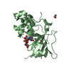
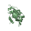
ジャーナル: PLoS Pathog / 年: 2015 タイトル: Dimerization-Induced Allosteric Changes of the Oxyanion-Hole Loop Activate the Pseudorabies Virus Assemblin pUL26N, a Herpesvirus Serine Protease. 著者: Martin Zühlsdorf / Sebastiaan Werten / Barbara G Klupp / Gottfried J Palm / Thomas C Mettenleiter / Winfried Hinrichs / 要旨: Herpesviruses encode a characteristic serine protease with a unique fold and an active site that comprises the unusual triad Ser-His-His. The protease is essential for viral replication and as such ...Herpesviruses encode a characteristic serine protease with a unique fold and an active site that comprises the unusual triad Ser-His-His. The protease is essential for viral replication and as such constitutes a promising drug target. In solution, a dynamic equilibrium exists between an inactive monomeric and an active dimeric form of the enzyme, which is believed to play a key regulatory role in the orchestration of proteolysis and capsid assembly. Currently available crystal structures of herpesvirus proteases correspond either to the dimeric state or to complexes with peptide mimetics that alter the dimerization interface. In contrast, the structure of the native monomeric state has remained elusive. Here, we present the three-dimensional structures of native monomeric, active dimeric, and diisopropyl fluorophosphate-inhibited dimeric protease derived from pseudorabies virus, an alphaherpesvirus of swine. These structures, solved by X-ray crystallography to respective resolutions of 2.05, 2.10 and 2.03 Å, allow a direct comparison of the main conformational states of the protease. In the dimeric form, a functional oxyanion hole is formed by a loop of 10 amino-acid residues encompassing two consecutive arginine residues (Arg136 and Arg137); both are strictly conserved throughout the herpesviruses. In the monomeric form, the top of the loop is shifted by approximately 11 Å, resulting in a complete disruption of the oxyanion hole and loss of activity. The dimerization-induced allosteric changes described here form the physical basis for the concentration-dependent activation of the protease, which is essential for proper virus replication. Small-angle X-ray scattering experiments confirmed a concentration-dependent equilibrium of monomeric and dimeric protease in solution.
履歴
登録
2014年4月4日
登録サイト: PDBE / 処理サイト: PDBE
改定 1.0
2015年5月20日
Provider: repository / タイプ: Initial release
改定 1.1
2015年7月15日
Group: Database references / Other / Structure summary
SHEET DETERMINATION METHOD: DSSP THE SHEETS PRESENTED AS "BA" IN EACH CHAIN ON SHEET RECORDS BELOW ... SHEET DETERMINATION METHOD: DSSP THE SHEETS PRESENTED AS "BA" IN EACH CHAIN ON SHEET RECORDS BELOW IS ACTUALLY AN 7-STRANDED BARREL THIS IS REPRESENTED BY A 8-STRANDED SHEET IN WHICH THE FIRST AND LAST STRANDS ARE IDENTICAL.
モノクロメーター: SI(111) / プロトコル: SINGLE WAVELENGTH / 単色(M)・ラウエ(L): M / 散乱光タイプ: x-ray
放射波長
波長: 0.91841 Å / 相対比: 1
反射
解像度: 2.53→80 Å / Num. obs: 18230 / % possible obs: 97.8 % / Observed criterion σ(I): -3 / 冗長度: 3.8 % / Biso Wilson estimate: 55.9 Å2 / Rmerge(I) obs: 0.08 / Net I/σ(I): 14.29
反射 シェル
解像度: 2.53→2.68 Å / 冗長度: 3.3 % / Rmerge(I) obs: 0.66 / Mean I/σ(I) obs: 2.06 / % possible all: 93.6
-
解析
ソフトウェア
名称
バージョン
分類
REFMAC
5.8.0069
精密化
XDS
データ削減
Aimless
データスケーリング
PHASER
位相決定
精密化
構造決定の手法: 分子置換 開始モデル: DIMERIC PRV PROTEASE, YET TO BE DEPOSITED 解像度: 2.53→79.94 Å / Cor.coef. Fo:Fc: 0.949 / Cor.coef. Fo:Fc free: 0.912 / SU B: 21.997 / SU ML: 0.232 / 交差検証法: THROUGHOUT / ESU R: 0.423 / ESU R Free: 0.279 / 立体化学のターゲット値: MAXIMUM LIKELIHOOD 詳細: HYDROGENS HAVE BEEN ADDED IN THE RIDING POSITIONS. VALUES WITH TLS ADDED
Rfactor
反射数
%反射
Selection details
Rfree
0.25215
878
4.8 %
RANDOM
Rwork
0.19819
-
-
-
obs
0.20078
17352
97.75 %
-
溶媒の処理
イオンプローブ半径: 0.8 Å / 減衰半径: 0.8 Å / VDWプローブ半径: 1.1 Å / 溶媒モデル: MASK
 ムービー
ムービー コントローラー


 データを開く
 基本情報
基本情報 要素
要素 キーワード
キーワード 機能・相同性情報
 SUID HERPESVIRUS 1 (ヘルペスウイルス)
 X線回折 /
  データ登録者
 引用
引用
 構造の表示
構造の表示 ダウンロードとリンク
 その他のダウンロード


 PDBj
PDBj 集合体
集合体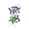


 分子量: 18.015 Da / 分子数: 49 / 由来タイプ: 天然 / 式: H2O
 試料調製
試料調製 解析
解析