 Movie
Movie Controller

[English] 日本語
 Yorodumi
- PDB-5llt: Plasmodium falciparum nicotinic acid mononucleotide adenylyltrans... -
+ Open data

- Basic information
| Entry | Database: PDB / ID: 5llt | ||||||
|---|---|---|---|---|---|---|---|
| Title | Plasmodium falciparum nicotinic acid mononucleotide adenylyltransferase complexed with NaAD | ||||||
 Components Components | Nicotinate-nucleotide adenylyltransferase | ||||||
 Keywords Keywords | TRANSFERASE / Nicotinic acid mononucleotide adenylyltransferase / NaMNAT / protein crystallography / Plasmodium falciparum / drug target / malaria / NAD metabolism | ||||||
| Function / homology |  Function and homology information Function and homology informationnicotinate-nucleotide adenylyltransferase / nicotinate-nucleotide adenylyltransferase activity / NAD+ biosynthetic process / ATP binding / cytoplasm Similarity search - Function | ||||||
| Biological species |  | ||||||
| Method |  X-RAY DIFFRACTION / SYNCHROTRON / MOLECULAR REPLACEMENT / Resolution: 2.2 Å X-RAY DIFFRACTION / SYNCHROTRON / MOLECULAR REPLACEMENT / Resolution: 2.2 Å | ||||||
 Authors Authors | Bathke, J. / Fritz-Wolf, K. / Brandstaedter, C. / Rahlfs, S. / Becker, K. | ||||||
| Funding support |  Germany, 1items Germany, 1items
| ||||||
 Citation Citation | Journal: J. Mol. Biol. / Year: 2016 Title: Structural and Functional Characterization of Plasmodium falciparum Nicotinic Acid Mononucleotide Adenylyltransferase. Authors: Bathke, J. / Fritz-Wolf, K. / Brandstadter, C. / Burkhardt, A. / Jortzik, E. / Rahlfs, S. / Becker, K. | ||||||
| History |
|
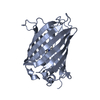
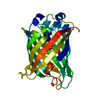
- Structure visualization
| Structure viewer | Molecule: MolmilJmol/JSmol |
|---|
- Downloads & links
-Download
| PDBx/mmCIF format | 5llt.cif.gz | 104.6 KB | Display | PDBx/mmCIF format |
|---|---|---|---|---|
| PDB format | pdb5llt.ent.gz | 80.9 KB | Display | PDB format |
| PDBx/mmJSON format | 5llt.json.gz | Tree view | PDBx/mmJSON format | |
| Others |  Other downloads Other downloads |
-Validation report
| Arichive directory | https://data.pdbj.org/pub/pdb/validation_reports/ll/5lltftp://data.pdbj.org/pub/pdb/validation_reports/ll/5llt | HTTPS FTP |
|---|
-Related structure data
| Related structure data |  5lm3C  2h29S S: Starting model for refinement C: citing same article ( |
|---|---|
| Similar structure data |


-Links
 PDBj
PDBj- Assembly
| Deposited unit | 
| ||||||||
|---|---|---|---|---|---|---|---|---|---|
| 1 | 
| ||||||||
| 2 | 
| ||||||||
| Unit cell |
|
-Components
| #1: Protein | Mass: 25445.338 Da / Num. of mol.: 2 Source method: isolated from a genetically manipulated source Source: (gene. exp.) Strain: isolate 3D7 / Gene: PF3D7_1327600 / Production host:  References: UniProt: Q8IE38, nicotinate-nucleotide adenylyltransferase #2: Chemical |   Mass: 665.418 Da / Num. of mol.: 2 / Source method: obtained synthetically / Formula: C21H27N6O15P2 Mass: 665.418 Da / Num. of mol.: 2 / Source method: obtained synthetically / Formula: C21H27N6O15P2#3: Chemical |   Mass: 106.120 Da / Num. of mol.: 2 / Source method: obtained synthetically / Formula: C4H10O3 Mass: 106.120 Da / Num. of mol.: 2 / Source method: obtained synthetically / Formula: C4H10O3#4: Chemical | ChemComp-EDO /   Mass: 62.068 Da / Num. of mol.: 9 / Source method: obtained synthetically / Formula: C2H6O2 Mass: 62.068 Da / Num. of mol.: 9 / Source method: obtained synthetically / Formula: C2H6O2#5: Water | ChemComp-HOH / |  Mass: 18.015 Da / Num. of mol.: 154 / Source method: isolated from a natural source / Formula: H2O Mass: 18.015 Da / Num. of mol.: 154 / Source method: isolated from a natural source / Formula: H2O |
|---|
-Experimental details
-Experiment
| Experiment | Method: X-RAY DIFFRACTION / Number of used crystals: 1 |
|---|
- Sample preparation
| Crystal | Density Matthews: 2.29 Å3/Da / Density % sol: 46.35 % |
|---|---|
| Crystal grow | Temperature: 300 K / Method: vapor diffusion, sitting drop / Details: 16 mg/ml, 0.1 M bicine, pH 8.2 and 18% PEG 6000. |
-Data collection
| Diffraction | Mean temperature: 100 K |
|---|---|
| Diffraction source | Source: SYNCHROTRON / Site: SLS  / Beamline: X10SA / Wavelength: 1 Å / Beamline: X10SA / Wavelength: 1 Å |
| Detector | Type: DECTRIS PILATUS3 6M / Detector: PIXEL / Date: Apr 3, 2014 |
| Radiation | Protocol: SINGLE WAVELENGTH / Monochromatic (M) / Laue (L): M / Scattering type: x-ray |
| Radiation wavelength | Wavelength: 1 Å / Relative weight: 1 |
| Reflection | Resolution: 2.2→40 Å / Num. obs: 19100 / % possible obs: 83.3 % / Redundancy: 1.7 % / CC1/2: 0.998 / Rmerge(I) obs: 0.048 / Rsym value: 0.034 / Net I/av σ(I): 14.17 / Net I/σ(I): 14.1 |
| Reflection shell | Resolution: 2.2→2.26 Å / Redundancy: 1.65 % / Rmerge(I) obs: 0.2 / Mean I/σ(I) obs: 4.97 / CC1/2: 0.969 / % possible all: 41.3 |
- Processing
| Software |
| |||||||||||||||||||||||||||||||||||||||||||||||||||||||||||||||
|---|---|---|---|---|---|---|---|---|---|---|---|---|---|---|---|---|---|---|---|---|---|---|---|---|---|---|---|---|---|---|---|---|---|---|---|---|---|---|---|---|---|---|---|---|---|---|---|---|---|---|---|---|---|---|---|---|---|---|---|---|---|---|---|---|
| Refinement | Method to determine structure: MOLECULAR REPLACEMENT Starting model: 2H29 Resolution: 2.2→39.603 Å / SU ML: 0.29 / Cross valid method: FREE R-VALUE / σ(F): 2.03 / Phase error: 27.88 / Stereochemistry target values: ML
| |||||||||||||||||||||||||||||||||||||||||||||||||||||||||||||||
| Solvent computation | Shrinkage radii: 0.9 Å / VDW probe radii: 1.11 Å / Solvent model: FLAT BULK SOLVENT MODEL | |||||||||||||||||||||||||||||||||||||||||||||||||||||||||||||||
| Refinement step | Cycle: LAST / Resolution: 2.2→39.603 Å
| |||||||||||||||||||||||||||||||||||||||||||||||||||||||||||||||
| Refine LS restraints |
| |||||||||||||||||||||||||||||||||||||||||||||||||||||||||||||||
| LS refinement shell |
|
