 Movie
Movie Controller

[English] 日本語
 Yorodumi
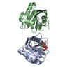
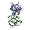
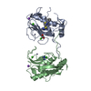
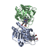
- PDB-4cn9: structure of proximal thread matrix protein 1 (PTMP1) from the mu... -
+ Open data

- Basic information
| Entry | Database: PDB / ID: 4cn9 | ||||||
|---|---|---|---|---|---|---|---|
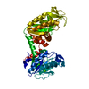
| Title | structure of proximal thread matrix protein 1 (PTMP1) from the mussel byssus with zinc occupied MIDAS motif | ||||||
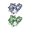
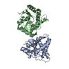
 Components Components | PROXIMAL THREAD MATRIX PROTEIN 1 | ||||||
 Keywords Keywords | STRUCTURAL PROTEIN | ||||||
| Function / homology |  Function and homology information Function and homology information | ||||||
| Biological species |  MYTILUS GALLOPROVINCIALIS (Mediterranean mussel) MYTILUS GALLOPROVINCIALIS (Mediterranean mussel) | ||||||
| Method |  X-RAY DIFFRACTION / SYNCHROTRON / MOLECULAR REPLACEMENT / Resolution: 1.9 Å X-RAY DIFFRACTION / SYNCHROTRON / MOLECULAR REPLACEMENT / Resolution: 1.9 Å | ||||||
 Authors Authors | Gertz, M. / Suhre, M.H. / Scheibel, T. / Steegborn, C. | ||||||
 Citation Citation | Journal: Nat.Commun. / Year: 2014 Title: Structural and Functional Features of a Collagen-Binding Matrix Protein from the Mussel Byssus. Authors: Suhre, M.H. / Gertz, M. / Steegborn, C. / Scheibel, T. | ||||||
| History |
|
- Structure visualization
| Structure viewer | Molecule: MolmilJmol/JSmol |
|---|
- Downloads & links
-Download
| PDBx/mmCIF format | 4cn9.cif.gz | 303.5 KB | Display | PDBx/mmCIF format |
|---|---|---|---|---|
| PDB format | pdb4cn9.ent.gz | 248.7 KB | Display | PDB format |
| PDBx/mmJSON format | 4cn9.json.gz | Tree view | PDBx/mmJSON format | |
| Others |  Other downloads Other downloads |
-Validation report
| Arichive directory | https://data.pdbj.org/pub/pdb/validation_reports/cn/4cn9ftp://data.pdbj.org/pub/pdb/validation_reports/cn/4cn9 | HTTPS FTP |
|---|
-Related structure data
| Related structure data |  4cn8C 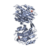 4cnbSC C: citing same article ( S: Starting model for refinement |
|---|---|
| Similar structure data |


-Links
 PDBj
PDBj


- Assembly
| Deposited unit | 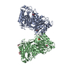
| ||||||||
|---|---|---|---|---|---|---|---|---|---|
| 1 | 
| ||||||||
| 2 | 
| ||||||||
| Unit cell |
| ||||||||
| Noncrystallographic symmetry (NCS) | NCS oper: (Code: given Matrix: (0.917, -0.000264, 0.3988), Vector: |
-Components
-Protein , 1 types, 2 molecules AB
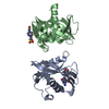
| #1: Protein | Mass: 48970.336 Da / Num. of mol.: 2 Source method: isolated from a genetically manipulated source Source: (gene. exp.) MYTILUS GALLOPROVINCIALIS (Mediterranean mussel)Production host:  |
|---|
-Non-polymers , 5 types, 411 molecules 


| #2: Chemical | ChemComp-ZN /  Mass: 65.409 Da / Num. of mol.: 4 / Source method: obtained synthetically / Formula: Zn Mass: 65.409 Da / Num. of mol.: 4 / Source method: obtained synthetically / Formula: Zn#3: Chemical |  Mass: 62.068 Da / Num. of mol.: 3 / Source method: obtained synthetically / Formula: C2H6O2 Mass: 62.068 Da / Num. of mol.: 3 / Source method: obtained synthetically / Formula: C2H6O2#4: Chemical | ChemComp-GOL /  Mass: 92.094 Da / Num. of mol.: 4 / Source method: obtained synthetically / Formula: C3H8O3 Mass: 92.094 Da / Num. of mol.: 4 / Source method: obtained synthetically / Formula: C3H8O3#5: Chemical |  Mass: 96.063 Da / Num. of mol.: 2 / Source method: obtained synthetically / Formula: SO4 Mass: 96.063 Da / Num. of mol.: 2 / Source method: obtained synthetically / Formula: SO4#6: Water | ChemComp-HOH / | Mass: 18.015 Da / Num. of mol.: 398 / Source method: isolated from a natural source / Formula: H2O |
|---|
-Details
| Has protein modification | Y |
|---|
-Experimental details
-Experiment
| Experiment | Method: X-RAY DIFFRACTION / Number of used crystals: 1 |
|---|
- Sample preparation
| Crystal | Density Matthews: 2.37 Å3/Da / Density % sol: 48.16 % / Description: NONE |
|---|---|
| Crystal grow | pH: 4.6 Details: 28% PEG2000-MME, 0.2 M AMMONIUM SULFATE, 0.1 M SODIUM ACETATE PH 4.6 |
-Data collection
| Diffraction | Mean temperature: 100 K |
|---|---|
| Diffraction source | Source: SYNCHROTRON / Site: BESSY  / Beamline: 14.1 / Wavelength: 1.283 / Beamline: 14.1 / Wavelength: 1.283 |
| Detector | Type: MARMOSAIC 225 mm CCD / Detector: CCD / Date: Nov 14, 2012 / Details: COLLIMATOR |
| Radiation | Monochromator: SI(111) MONOCHROMATOR / Protocol: SINGLE WAVELENGTH / Monochromatic (M) / Laue (L): M / Scattering type: x-ray |
| Radiation wavelength | Wavelength: 1.283 Å / Relative weight: 1 |
| Reflection | Resolution: 1.9→43.6 Å / Num. obs: 72516 / % possible obs: 99.9 % / Observed criterion σ(I): -3 / Redundancy: 4.1 % / Biso Wilson estimate: 23.39 Å2 / Rmerge(I) obs: 0.1 / Net I/σ(I): 11 |
| Reflection shell | Resolution: 1.9→2 Å / Redundancy: 4.1 % / Rmerge(I) obs: 1.03 / Mean I/σ(I) obs: 1.5 / % possible all: 99.9 |
- Processing
| Software |
| |||||||||||||||||||||||||||||||||||||||||||||||||||||||||||||||||||||||||||||||||||||||||||||||||||||||||||||||||||||||||||||||||||||||||||||||||||||||||||||||||||||||||||||||||||||||||||||
|---|---|---|---|---|---|---|---|---|---|---|---|---|---|---|---|---|---|---|---|---|---|---|---|---|---|---|---|---|---|---|---|---|---|---|---|---|---|---|---|---|---|---|---|---|---|---|---|---|---|---|---|---|---|---|---|---|---|---|---|---|---|---|---|---|---|---|---|---|---|---|---|---|---|---|---|---|---|---|---|---|---|---|---|---|---|---|---|---|---|---|---|---|---|---|---|---|---|---|---|---|---|---|---|---|---|---|---|---|---|---|---|---|---|---|---|---|---|---|---|---|---|---|---|---|---|---|---|---|---|---|---|---|---|---|---|---|---|---|---|---|---|---|---|---|---|---|---|---|---|---|---|---|---|---|---|---|---|---|---|---|---|---|---|---|---|---|---|---|---|---|---|---|---|---|---|---|---|---|---|---|---|---|---|---|---|---|---|---|---|---|
| Refinement | Method to determine structure: MOLECULAR REPLACEMENT Starting model: PDB ENTRY 4CNB Resolution: 1.9→43.559 Å / SU ML: 0.2 / σ(F): 1.99 / Phase error: 20.81 / Stereochemistry target values: ML
| |||||||||||||||||||||||||||||||||||||||||||||||||||||||||||||||||||||||||||||||||||||||||||||||||||||||||||||||||||||||||||||||||||||||||||||||||||||||||||||||||||||||||||||||||||||||||||||
| Solvent computation | Shrinkage radii: 0.9 Å / VDW probe radii: 1.11 Å / Solvent model: FLAT BULK SOLVENT MODEL | |||||||||||||||||||||||||||||||||||||||||||||||||||||||||||||||||||||||||||||||||||||||||||||||||||||||||||||||||||||||||||||||||||||||||||||||||||||||||||||||||||||||||||||||||||||||||||||
| Displacement parameters | Biso mean: 30.2 Å2 | |||||||||||||||||||||||||||||||||||||||||||||||||||||||||||||||||||||||||||||||||||||||||||||||||||||||||||||||||||||||||||||||||||||||||||||||||||||||||||||||||||||||||||||||||||||||||||||
| Refinement step | Cycle: LAST / Resolution: 1.9→43.559 Å
| |||||||||||||||||||||||||||||||||||||||||||||||||||||||||||||||||||||||||||||||||||||||||||||||||||||||||||||||||||||||||||||||||||||||||||||||||||||||||||||||||||||||||||||||||||||||||||||
| Refine LS restraints |
| |||||||||||||||||||||||||||||||||||||||||||||||||||||||||||||||||||||||||||||||||||||||||||||||||||||||||||||||||||||||||||||||||||||||||||||||||||||||||||||||||||||||||||||||||||||||||||||
| LS refinement shell |
|
