 Movie
Movie Controller

[English] 日本語
 Yorodumi
- PDB-4cgy: Crystal structure of the human topoisomerase III alpha-RMI1 complex -
+ Open data

- Basic information
| Entry | Database: PDB / ID: 4cgy | ||||||
|---|---|---|---|---|---|---|---|
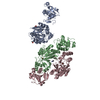
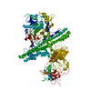
| Title | Crystal structure of the human topoisomerase III alpha-RMI1 complex | ||||||
 Components Components |
| ||||||
 Keywords Keywords | DNA REPLICATION/ISOMERASE / DNA REPLICATION-ISOMERASE COMPLEX / DOUBLE HOLLIDAY JUNCTION DISSOLUTION / DECATENATION / MINIMAL DISSOLVASOME | ||||||
| Function / homology |  Function and homology information Function and homology informationRecQ family helicase-topoisomerase III complex / reduction of food intake in response to dietary excess / resolution of DNA recombination intermediates / chromosome separation / DNA topoisomerase / DNA topoisomerase type I (single strand cut, ATP-independent) activity / mitochondrial DNA metabolic process / resolution of meiotic recombination intermediates / Strand-asynchronous mitochondrial DNA replication / Impaired BRCA2 binding to PALB2 ...RecQ family helicase-topoisomerase III complex / reduction of food intake in response to dietary excess / resolution of DNA recombination intermediates / chromosome separation / DNA topoisomerase / DNA topoisomerase type I (single strand cut, ATP-independent) activity / mitochondrial DNA metabolic process / resolution of meiotic recombination intermediates / Strand-asynchronous mitochondrial DNA replication / Impaired BRCA2 binding to PALB2 / HDR through Single Strand Annealing (SSA) / Homologous DNA Pairing and Strand Exchange / Defective homologous recombination repair (HRR) due to BRCA1 loss of function / Defective HDR through Homologous Recombination Repair (HRR) due to PALB2 loss of BRCA1 binding function / Defective HDR through Homologous Recombination Repair (HRR) due to PALB2 loss of BRCA2/RAD51/RAD51C binding function / Resolution of D-loop Structures through Synthesis-Dependent Strand Annealing (SDSA) / Resolution of D-loop Structures through Holliday Junction Intermediates / Impaired BRCA2 binding to RAD51 / DNA topological change / Presynaptic phase of homologous DNA pairing and strand exchange / response to glucose / meiotic cell cycle / PML body / G2/M DNA damage checkpoint / double-strand break repair via homologous recombination / Meiotic recombination / HDR through Homologous Recombination (HRR) / multicellular organism growth / glucose homeostasis / single-stranded DNA binding / Processing of DNA double-strand break ends / DNA recombination / Regulation of TP53 Activity through Phosphorylation / DNA replication / nuclear body / mitochondrial matrix / nucleotide binding / DNA repair / mitochondrion / DNA binding / zinc ion binding / nucleoplasm / nucleus Similarity search - Function | ||||||
| Biological species |  HOMO SAPIENS (human) HOMO SAPIENS (human) | ||||||
| Method |  X-RAY DIFFRACTION / SYNCHROTRON / MOLECULAR REPLACEMENT / Resolution: 2.85 Å X-RAY DIFFRACTION / SYNCHROTRON / MOLECULAR REPLACEMENT / Resolution: 2.85 Å | ||||||
 Authors Authors | Bocquet, N. / Bunker, R.D. / Thoma, N.H. | ||||||
 Citation Citation | Journal: Nat.Struct.Mol.Biol. / Year: 2014 Title: Structural and Mechanistic Insight Into Holliday-Junction Dissolution by Topoisomerase Iiialpha and Rmi1 Authors: Bocquet, N. / Bizard, A.H. / Abdulrahman, W. / Larsen, N.B. / Faty, M. / Cavadini, S. / Bunker, R.D. / Kowalczykowski, S.C. / Cejka, P. / Hickson, I.D. / Thoma, N.H. | ||||||
| History |
|
- Structure visualization
| Structure viewer | Molecule: MolmilJmol/JSmol |
|---|
- Downloads & links
-Download
| PDBx/mmCIF format | 4cgy.cif.gz | 343.9 KB | Display | PDBx/mmCIF format |
|---|---|---|---|---|
| PDB format | pdb4cgy.ent.gz | 279.8 KB | Display | PDB format |
| PDBx/mmJSON format | 4cgy.json.gz | Tree view | PDBx/mmJSON format | |
| Others |  Other downloads Other downloads |
-Validation report
| Arichive directory | https://data.pdbj.org/pub/pdb/validation_reports/cg/4cgyftp://data.pdbj.org/pub/pdb/validation_reports/cg/4cgy | HTTPS FTP |
|---|
-Related structure data
| Related structure data |  4chtC  1eclS  3nbiS S: Starting model for refinement C: citing same article ( |
|---|---|
| Similar structure data |


-Links
 PDBj
PDBj


- Assembly
| Deposited unit | 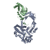
| ||||||||
|---|---|---|---|---|---|---|---|---|---|
| 1 |
| ||||||||
| Unit cell |
|
-Components
| #1: Protein | Mass: 86584.641 Da / Num. of mol.: 1 / Fragment: RESIDUES 2-753 Source method: isolated from a genetically manipulated source Source: (gene. exp.) HOMO SAPIENS (human) / Cell line (production host): High Five / Production host:  TRICHOPLUSIA NI (cabbage looper) / References: UniProt: Q13472, EC: 5.99.1.2 TRICHOPLUSIA NI (cabbage looper) / References: UniProt: Q13472, EC: 5.99.1.2 |
|---|---|
| #2: Protein | Mass: 24644.457 Da / Num. of mol.: 1 / Fragment: RESIDUES 1-219 Source method: isolated from a genetically manipulated source Source: (gene. exp.) HOMO SAPIENS (human) / Cell line (production host): High Five / Production host: TRICHOPLUSIA NI (cabbage looper) / References: UniProt: Q9H9A7 |
| #3: Chemical | ChemComp-MG /   Mass: 24.305 Da / Num. of mol.: 1 / Source method: obtained synthetically / Formula: Mg Mass: 24.305 Da / Num. of mol.: 1 / Source method: obtained synthetically / Formula: Mg |
| #4: Water | ChemComp-HOH /  Mass: 18.015 Da / Num. of mol.: 44 / Source method: isolated from a natural source / Formula: H2O Mass: 18.015 Da / Num. of mol.: 44 / Source method: isolated from a natural source / Formula: H2O |
-Experimental details
-Experiment
| Experiment | Method: X-RAY DIFFRACTION / Number of used crystals: 1 |
|---|
- Sample preparation
| Crystal | Density Matthews: 5.4 Å3/Da / Density % sol: 77 % / Description: NONE |
|---|---|
| Crystal grow | pH: 7 Details: 8-12% (W/V) PEG 2000, 100 MM TRIS-HCL PH 7.0, 200 MM MGCL2 |
-Data collection
| Diffraction | Mean temperature: 10 K |
|---|---|
| Diffraction source | Source: SYNCHROTRON / Site: SLS  / Beamline: X10SA / Wavelength: 1 / Beamline: X10SA / Wavelength: 1 |
| Detector | Type: DECTRIS PILATUS 6M / Detector: PIXEL / Date: Sep 19, 2010 / Details: DYNAMICALLY BENDABLE MIRROR |
| Radiation | Monochromator: SI(111) / Protocol: SINGLE WAVELENGTH / Monochromatic (M) / Laue (L): M / Scattering type: x-ray |
| Radiation wavelength | Wavelength: 1 Å / Relative weight: 1 |
| Reflection | Resolution: 2.85→62.78 Å / Num. obs: 41328 / % possible obs: 100 % / Observed criterion σ(I): -3 / Redundancy: 100 % / Biso Wilson estimate: 93.75 Å2 / Rmerge(I) obs: 0.15 / Net I/σ(I): 16.7 |
| Reflection shell | Resolution: 2.85→2.97 Å / Redundancy: 14.7 % / Mean I/σ(I) obs: 2.2 / % possible all: 100 |
- Processing
| Software |
| ||||||||||||||||||||||||||||||||||||||||||||||||||||||||||||||||||||||||||||||||||||||||||||||||||||||||||||||||||
|---|---|---|---|---|---|---|---|---|---|---|---|---|---|---|---|---|---|---|---|---|---|---|---|---|---|---|---|---|---|---|---|---|---|---|---|---|---|---|---|---|---|---|---|---|---|---|---|---|---|---|---|---|---|---|---|---|---|---|---|---|---|---|---|---|---|---|---|---|---|---|---|---|---|---|---|---|---|---|---|---|---|---|---|---|---|---|---|---|---|---|---|---|---|---|---|---|---|---|---|---|---|---|---|---|---|---|---|---|---|---|---|---|---|---|---|
| Refinement | Method to determine structure: MOLECULAR REPLACEMENT Starting model: PDB ENTRIES 1ECL AND 3NBI Resolution: 2.85→65.49 Å / Cor.coef. Fo:Fc: 0.9298 / Cor.coef. Fo:Fc free: 0.9116 / SU R Cruickshank DPI: 0.374 / Cross valid method: THROUGHOUT / σ(F): 0 / SU R Blow DPI: 0.376 / SU Rfree Blow DPI: 0.26 / SU Rfree Cruickshank DPI: 0.263
| ||||||||||||||||||||||||||||||||||||||||||||||||||||||||||||||||||||||||||||||||||||||||||||||||||||||||||||||||||
| Displacement parameters | Biso mean: 80.28 Å2
| ||||||||||||||||||||||||||||||||||||||||||||||||||||||||||||||||||||||||||||||||||||||||||||||||||||||||||||||||||
| Refine analyze | Luzzati coordinate error obs: 0.439 Å | ||||||||||||||||||||||||||||||||||||||||||||||||||||||||||||||||||||||||||||||||||||||||||||||||||||||||||||||||||
| Refinement step | Cycle: LAST / Resolution: 2.85→65.49 Å
| ||||||||||||||||||||||||||||||||||||||||||||||||||||||||||||||||||||||||||||||||||||||||||||||||||||||||||||||||||
| Refine LS restraints |
| ||||||||||||||||||||||||||||||||||||||||||||||||||||||||||||||||||||||||||||||||||||||||||||||||||||||||||||||||||
| LS refinement shell | Resolution: 2.85→2.92 Å / Total num. of bins used: 20
| ||||||||||||||||||||||||||||||||||||||||||||||||||||||||||||||||||||||||||||||||||||||||||||||||||||||||||||||||||
| Refinement TLS params. | Method: refined / Refine-ID: X-RAY DIFFRACTION
| ||||||||||||||||||||||||||||||||||||||||||||||||||||||||||||||||||||||||||||||||||||||||||||||||||||||||||||||||||
| Refinement TLS group |
|
