 Movie
Movie Controller


+ Open data

- Basic information
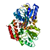
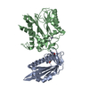
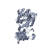
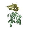
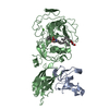
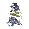
| Entry | Database: PDB / ID: 6ob5 | |||||||||
|---|---|---|---|---|---|---|---|---|---|---|
| Title | Computationally-designed, modular sense/response system (S3-2D) | |||||||||
 Components Components |
| |||||||||
 Keywords Keywords | SUGAR BINDING PROTEIN / computational protein design / chemically-induced dimerization / biosensor / Rosetta | |||||||||
| Function / homology |  Function and homology information Function and homology informationdetection of maltose stimulus / maltose transport complex / carbohydrate transport / carbohydrate transmembrane transporter activity / maltose binding / maltose transport / maltodextrin transmembrane transport / ATP-binding cassette (ABC) transporter complex, substrate-binding subunit-containing / ATP-binding cassette (ABC) transporter complex / cell chemotaxis ...detection of maltose stimulus / maltose transport complex / carbohydrate transport / carbohydrate transmembrane transporter activity / maltose binding / maltose transport / maltodextrin transmembrane transport / ATP-binding cassette (ABC) transporter complex, substrate-binding subunit-containing / ATP-binding cassette (ABC) transporter complex / cell chemotaxis / outer membrane-bounded periplasmic space / periplasmic space / DNA damage response / membrane Similarity search - Function | |||||||||
| Biological species |  unidentified (others) | |||||||||
| Method |  X-RAY DIFFRACTION / SYNCHROTRON / MOLECULAR REPLACEMENT / Resolution: 2.208 Å X-RAY DIFFRACTION / SYNCHROTRON / MOLECULAR REPLACEMENT / Resolution: 2.208 Å | |||||||||
 Authors Authors | Thompson, M.C. / Glasgow, A.A. / Huang, Y.M. / Fraser, J.S. / Kortemme, T. | |||||||||
| Funding support |  United States, 1items United States, 1items
| |||||||||
 Citation Citation | Journal: Science / Year: 2019 Title: Computational design of a modular protein sense-response system. Authors: Glasgow, A.A. / Huang, Y.M. / Mandell, D.J. / Thompson, M. / Ritterson, R. / Loshbaugh, A.L. / Pellegrino, J. / Krivacic, C. / Pache, R.A. / Barlow, K.A. / Ollikainen, N. / Jeon, D. / Kelly, ...Authors: Glasgow, A.A. / Huang, Y.M. / Mandell, D.J. / Thompson, M. / Ritterson, R. / Loshbaugh, A.L. / Pellegrino, J. / Krivacic, C. / Pache, R.A. / Barlow, K.A. / Ollikainen, N. / Jeon, D. / Kelly, M.J.S. / Fraser, J.S. / Kortemme, T. | |||||||||
| History |
|
- Structure visualization
| Structure viewer | Molecule: MolmilJmol/JSmol |
|---|
- Downloads & links
-Download
| PDBx/mmCIF format | 6ob5.cif.gz | 374.6 KB | Display | PDBx/mmCIF format |
|---|---|---|---|---|
| PDB format | pdb6ob5.ent.gz | 307.1 KB | Display | PDB format |
| PDBx/mmJSON format | 6ob5.json.gz | Tree view | PDBx/mmJSON format | |
| Others |  Other downloads Other downloads |
-Validation report
| Arichive directory | https://data.pdbj.org/pub/pdb/validation_reports/ob/6ob5ftp://data.pdbj.org/pub/pdb/validation_reports/ob/6ob5 | HTTPS FTP |
|---|
-Related structure data


-Links
 PDBj
PDBj


- Assembly
| Deposited unit | 
| ||||||||
|---|---|---|---|---|---|---|---|---|---|
| 1 | 
| ||||||||
| 2 | 
| ||||||||
| Unit cell |
|
-Components
| #1: Protein | Mass: 40825.281 Da / Num. of mol.: 2 Source method: isolated from a genetically manipulated source Source: (gene. exp.) #2: Protein | Mass: 17778.896 Da / Num. of mol.: 2 Source method: isolated from a genetically manipulated source Details: computationally-designed sequence / Source: (gene. exp.) unidentified (others) / Variant: S3-2D / Production host: #3: Polysaccharide |   Source method: isolated from a genetically manipulated source Details: oligosaccharide / References: alpha-maltose #4: Chemical | ChemComp-FPP / | 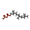  Mass: 382.326 Da / Num. of mol.: 1 / Source method: isolated from a natural source / Formula: C15H28O7P2 / Feature type: SUBJECT OF INVESTIGATION Mass: 382.326 Da / Num. of mol.: 1 / Source method: isolated from a natural source / Formula: C15H28O7P2 / Feature type: SUBJECT OF INVESTIGATION#5: Water | ChemComp-HOH / |  Mass: 18.015 Da / Num. of mol.: 141 / Source method: isolated from a natural source / Formula: H2O Mass: 18.015 Da / Num. of mol.: 141 / Source method: isolated from a natural source / Formula: H2O |
|---|
-Experimental details
-Experiment
| Experiment | Method: X-RAY DIFFRACTION / Number of used crystals: 1 |
|---|
- Sample preparation
| Crystal | Density Matthews: 2.12 Å3/Da / Density % sol: 41.86 % / Description: Thin, flat plates. Commonly stacked. |
|---|---|
| Crystal grow | Temperature: 293 K / Method: vapor diffusion, hanging drop / pH: 8.7 Details: 0.1M Tris Buffer, 0.1M sodium chloride, and 32% PEG-6000 PH range: 8.5-8.9 |
-Data collection
| Diffraction | Mean temperature: 100 K / Serial crystal experiment: N |
|---|---|
| Diffraction source | Source: SYNCHROTRON / Site: ALS / Beamline: 8.3.1 / Wavelength: 1.12 Å |
| Detector | Type: ADSC QUANTUM 315r / Detector: CCD / Date: Apr 22, 2016 |
| Radiation | Monochromator: Si111 / Protocol: SINGLE WAVELENGTH / Monochromatic (M) / Laue (L): M / Scattering type: x-ray |
| Radiation wavelength | Wavelength: 1.12 Å / Relative weight: 1 |
| Reflection | Resolution: 2.2→95.47 Å / Num. obs: 45972 / % possible obs: 98.5 % / Redundancy: 3.7 % / CC1/2: 0.997 / Rpim(I) all: 0.033 / Net I/σ(I): 17.5 |
| Reflection shell | Resolution: 2.2→2.24 Å / Redundancy: 3.6 % / Mean I/σ(I) obs: 8.3 / Num. unique obs: 7746 / CC1/2: 0.966 / Rpim(I) all: 0.09 / % possible all: 91.5 |
- Processing
| Software |
| |||||||||||||||||||||||||||||||||||||||||||||||||||||||||||||||||||||||||||||||||||||||||||||||||||||||||||||||||||||||||||||||||||||||||||||||||||
|---|---|---|---|---|---|---|---|---|---|---|---|---|---|---|---|---|---|---|---|---|---|---|---|---|---|---|---|---|---|---|---|---|---|---|---|---|---|---|---|---|---|---|---|---|---|---|---|---|---|---|---|---|---|---|---|---|---|---|---|---|---|---|---|---|---|---|---|---|---|---|---|---|---|---|---|---|---|---|---|---|---|---|---|---|---|---|---|---|---|---|---|---|---|---|---|---|---|---|---|---|---|---|---|---|---|---|---|---|---|---|---|---|---|---|---|---|---|---|---|---|---|---|---|---|---|---|---|---|---|---|---|---|---|---|---|---|---|---|---|---|---|---|---|---|---|---|---|---|
| Refinement | Method to determine structure: MOLECULAR REPLACEMENT Starting model: 1SVX, 1FQD Resolution: 2.208→95.46 Å / SU ML: 0 / Cross valid method: THROUGHOUT / σ(F): 1.99 / Phase error: 32.71
| |||||||||||||||||||||||||||||||||||||||||||||||||||||||||||||||||||||||||||||||||||||||||||||||||||||||||||||||||||||||||||||||||||||||||||||||||||
| Solvent computation | Shrinkage radii: 0.9 Å / VDW probe radii: 1.11 Å | |||||||||||||||||||||||||||||||||||||||||||||||||||||||||||||||||||||||||||||||||||||||||||||||||||||||||||||||||||||||||||||||||||||||||||||||||||
| Refinement step | Cycle: LAST / Resolution: 2.208→95.46 Å
| |||||||||||||||||||||||||||||||||||||||||||||||||||||||||||||||||||||||||||||||||||||||||||||||||||||||||||||||||||||||||||||||||||||||||||||||||||
| Refine LS restraints |
| |||||||||||||||||||||||||||||||||||||||||||||||||||||||||||||||||||||||||||||||||||||||||||||||||||||||||||||||||||||||||||||||||||||||||||||||||||
| LS refinement shell |
|
