 Movie
Movie Controller


+ Open data

- Basic information
| Entry | Database: PDB / ID: 3ttk | ||||||
|---|---|---|---|---|---|---|---|
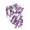
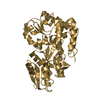
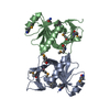
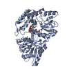
| Title | Crystal structure of apo-SpuD | ||||||
 Components Components | Polyamine transport protein | ||||||
 Keywords Keywords | TRANSPORT PROTEIN / polyamine receptor / polyamine binding | ||||||
| Function / homology |  Function and homology information Function and homology informationpolyamine transport / spermidine binding / putrescine binding / putrescine transport / outer membrane-bounded periplasmic space Similarity search - Function | ||||||
| Biological species |   Pseudomonas aeruginosa (bacteria) Pseudomonas aeruginosa (bacteria) | ||||||
| Method |  X-RAY DIFFRACTION / MOLECULAR REPLACEMENT / Resolution: 2.97 Å X-RAY DIFFRACTION / MOLECULAR REPLACEMENT / Resolution: 2.97 Å | ||||||
 Authors Authors | Wu, D.H. / Lim, S.C. / Song, H.W. | ||||||
 Citation Citation | Journal: J.Mol.Biol. / Year: 2012 Title: Structural Basis of Substrate Binding Specificity Revealed by the Crystal Structures of Polyamine Receptors SpuD and SpuE from Pseudomonas aeruginosa Authors: Wu, D.H. / Lim, S.C. / Dong, Y.H. / Wu, J.E. / Tao, F. / Zhou, L. / Zhang, L.H. / Song, H.W. | ||||||
| History |
|
- Structure visualization
| Structure viewer | Molecule: MolmilJmol/JSmol |
|---|
- Downloads & links
-Download
| PDBx/mmCIF format | 3ttk.cif.gz | 204.4 KB | Display | PDBx/mmCIF format |
|---|---|---|---|---|
| PDB format | pdb3ttk.ent.gz | 164.4 KB | Display | PDB format |
| PDBx/mmJSON format | 3ttk.json.gz | Tree view | PDBx/mmJSON format | |
| Others |  Other downloads Other downloads |
-Validation report
| Arichive directory | https://data.pdbj.org/pub/pdb/validation_reports/tt/3ttkftp://data.pdbj.org/pub/pdb/validation_reports/tt/3ttk | HTTPS FTP |
|---|
-Related structure data
| Related structure data |  3ttlC 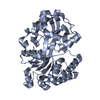 3ttmSC  3ttnC C: citing same article ( S: Starting model for refinement |
|---|---|
| Similar structure data |


-Links
 PDBj
PDBj


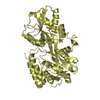
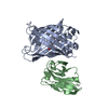
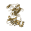
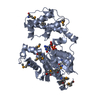
- Assembly
| Deposited unit | 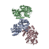
| ||||||||
|---|---|---|---|---|---|---|---|---|---|
| 1 | 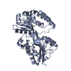
| ||||||||
| 2 | 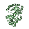
| ||||||||
| 3 | 
| ||||||||
| Unit cell |
|
-Components
| #1: Protein | Mass: 38380.617 Da / Num. of mol.: 3 / Fragment: UNP residues 26-365 Source method: isolated from a genetically manipulated source Source: (gene. exp.) Pseudomonas aeruginosa (bacteria) / Gene: spuD / Production host: #2: Water | ChemComp-HOH / |  Mass: 18.015 Da / Num. of mol.: 132 / Source method: isolated from a natural source / Formula: H2O Mass: 18.015 Da / Num. of mol.: 132 / Source method: isolated from a natural source / Formula: H2OHas protein modification | Y | |
|---|
-Experimental details
-Experiment
| Experiment | Method: X-RAY DIFFRACTION / Number of used crystals: 1 |
|---|
- Sample preparation
| Crystal | Density Matthews: 2.58 Å3/Da / Density % sol: 52.38 % |
|---|---|
| Crystal grow | Temperature: 288 K / Method: vapor diffusion, hanging drop / pH: 6.5 Details: 25% PEG 3350, 0.1M Mes, pH 6.5, VAPOR DIFFUSION, HANGING DROP, temperature 288K |
-Data collection
| Diffraction | Mean temperature: 100 K |
|---|---|
| Diffraction source | Source: ROTATING ANODE / Type: RIGAKU / Wavelength: 1.5418 Å |
| Detector | Type: RIGAKU / Detector: IMAGE PLATE / Date: Sep 15, 2009 |
| Radiation | Monochromator: Osmic mirrors / Protocol: SINGLE WAVELENGTH / Monochromatic (M) / Laue (L): M / Scattering type: x-ray |
| Radiation wavelength | Wavelength: 1.5418 Å / Relative weight: 1 |
| Reflection | Resolution: 2.9→20 Å / Num. all: 23272 / Num. obs: 23272 / % possible obs: 84.9 % / Observed criterion σ(F): 2 / Observed criterion σ(I): 2 |
| Reflection shell | Resolution: 2.9→3.06 Å / % possible all: 80.9 |
- Processing
| Software |
| |||||||||||||||||||||||||||||||||||||||||||||||||||||||||||||||||
|---|---|---|---|---|---|---|---|---|---|---|---|---|---|---|---|---|---|---|---|---|---|---|---|---|---|---|---|---|---|---|---|---|---|---|---|---|---|---|---|---|---|---|---|---|---|---|---|---|---|---|---|---|---|---|---|---|---|---|---|---|---|---|---|---|---|---|
| Refinement | Method to determine structure: MOLECULAR REPLACEMENT Starting model: 3TTM Resolution: 2.97→20 Å / Cor.coef. Fo:Fc: 0.92 / Cor.coef. Fo:Fc free: 0.853 / SU B: 20.282 / SU ML: 0.379 / Cross valid method: THROUGHOUT / σ(F): 2 / ESU R Free: 0.577 / Stereochemistry target values: MAXIMUM LIKELIHOOD / Details: HYDROGENS HAVE BEEN ADDED IN THE RIDING POSITIONS
| |||||||||||||||||||||||||||||||||||||||||||||||||||||||||||||||||
| Solvent computation | Ion probe radii: 0.8 Å / Shrinkage radii: 0.8 Å / VDW probe radii: 1.2 Å / Solvent model: MASK | |||||||||||||||||||||||||||||||||||||||||||||||||||||||||||||||||
| Displacement parameters | Biso mean: 39.859 Å2
| |||||||||||||||||||||||||||||||||||||||||||||||||||||||||||||||||
| Refinement step | Cycle: LAST / Resolution: 2.97→20 Å
| |||||||||||||||||||||||||||||||||||||||||||||||||||||||||||||||||
| Refine LS restraints |
| |||||||||||||||||||||||||||||||||||||||||||||||||||||||||||||||||
| LS refinement shell | Resolution: 2.97→3.045 Å / Total num. of bins used: 20
|
