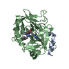
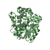
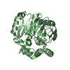
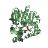
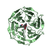
Entry Database : PDB / ID : 4ccwTitle Crystal structure of naproxen esterase (carboxylesterase NP) from Bacillus subtilis CARBOXYL ESTERASE NP Keywords Function / homology Function Domain/homology Component
/ / / / / / / / Biological species BACILLUS SUBTILIS (bacteria)Method / / / Resolution : 1.75 Å Authors Rozeboom, H.J. / Godinho, L.F. / Nardini, M. / Quax, W.J. / Dijkstra, B.W. Journal : Biochim.Biophys.Acta / Year : 2014Title : Crystal Structures of Two Bacillus Carboxylesterases with Different Enantioselectivities.Authors : Rozeboom, H.J. / Godinho, L.F. / Nardini, M. / Quax, W.J. / Dijkstra, B.W. History Deposition Oct 29, 2013 Deposition site / Processing site Revision 1.0 Jan 22, 2014 Provider / Type Revision 1.1 Aug 13, 2014 Group Revision 1.2 Apr 24, 2019 Group / Experimental preparation / OtherCategory exptl_crystal_grow / pdbx_database_proc ... exptl_crystal_grow / pdbx_database_proc / pdbx_database_status / struct_biol Item / _pdbx_database_status.recvd_author_approvalRevision 1.3 Jul 17, 2019 Group / Category / Item Revision 1.4 Dec 20, 2023 Group Data collection / Database references ... Data collection / Database references / Derived calculations / Other / Refinement description Category chem_comp_atom / chem_comp_bond ... chem_comp_atom / chem_comp_bond / database_2 / pdbx_database_status / pdbx_initial_refinement_model / struct_site Item _database_2.pdbx_DOI / _database_2.pdbx_database_accession ... _database_2.pdbx_DOI / _database_2.pdbx_database_accession / _pdbx_database_status.status_code_sf / _struct_site.pdbx_auth_asym_id / _struct_site.pdbx_auth_comp_id / _struct_site.pdbx_auth_seq_id
Show all Show less
 Movie
Movie Controller

 Yorodumi
 Open data
 Basic information
 Components
 Keywords
 Function and homology information
 X-RAY DIFFRACTION /
  Authors
 Citation
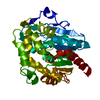
 Structure visualization
 Downloads & links
 Other downloads
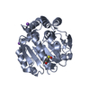
 PDBj
PDBj


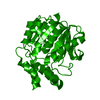
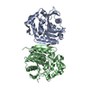
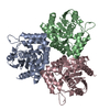
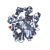
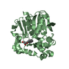
 Assembly
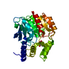
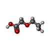
 Mass: 120.104 Da / Num. of mol.: 1 / Source method: obtained synthetically / Formula: C4H8O4
 Mass: 18.015 Da / Num. of mol.: 220 / Source method: isolated from a natural source / Formula: H2O
 Sample preparation
 / Beamline: BW7B / Wavelength: 1
  Processing