Resolution: 2.6→47.765 Å / SU ML: 0.82 / σ(F): 1.92 / Phase error: 27.49 / Stereochemistry target values: ML Details: PROTONS WERE MODELLED IN 'RIDING' POSITIONS. DISORDERED REGIONS WERE NOT MODELED
Rfactor
Num. reflection
% reflection
Rfree
0.2748
1095
5.1 %
Rwork
0.2425
-
-
obs
0.2441
21302
99.93 %
Solvent computation
Shrinkage radii: 0.9 Å / VDW probe radii: 1.11 Å / Solvent model: FLAT BULK SOLVENT MODEL / Bsol: 59.307 Å2 / ksol: 0.314 e/Å3
Displacement parameters
Biso mean: 95.38 Å2
Baniso -1
Baniso -2
Baniso -3
1-
5.1754 Å2
0 Å2
0 Å2
2-
-
5.1754 Å2
0 Å2
3-
-
-
-10.3508 Å2
Refinement step
Cycle: LAST / Resolution: 2.6→47.765 Å
Protein
Nucleic acid
Ligand
Solvent
Total
Num. atoms
4211
0
1
13
4225
Refine LS restraints
Refine-ID
Type
Dev ideal
Number
X-RAY DIFFRACTION
f_bond_d
0.003
4293
X-RAY DIFFRACTION
f_angle_d
0.798
5828
X-RAY DIFFRACTION
f_dihedral_angle_d
12.403
1599
X-RAY DIFFRACTION
f_chiral_restr
0.042
686
X-RAY DIFFRACTION
f_plane_restr
0.003
745
LS refinement shell
Resolution (Å)
Rfactor Rfree
Num. reflection Rfree
Rfactor Rwork
Num. reflection Rwork
Refine-ID
% reflection obs (%)
2.6002-2.7185
0.3601
139
0.3365
2427
X-RAY DIFFRACTION
100
2.7185-2.8618
0.3698
141
0.3208
2452
X-RAY DIFFRACTION
100
2.8618-3.0411
0.3447
116
0.2952
2495
X-RAY DIFFRACTION
100
3.0411-3.2759
0.3128
144
0.2935
2470
X-RAY DIFFRACTION
100
3.2759-3.6054
0.3325
152
0.2779
2488
X-RAY DIFFRACTION
100
3.6054-4.1269
0.2673
142
0.2476
2527
X-RAY DIFFRACTION
100
4.1269-5.1984
0.2525
125
0.198
2599
X-RAY DIFFRACTION
100
5.1984-47.7732
0.2283
136
0.2246
2749
X-RAY DIFFRACTION
100
Refinement TLS params.
Method: refined / Refine-ID: X-RAY DIFFRACTION
ID
L11 (°2)
L12 (°2)
L13 (°2)
L22 (°2)
L23 (°2)
L33 (°2)
S11 (Å °)
S12 (Å °)
S13 (Å °)
S21 (Å °)
S22 (Å °)
S23 (Å °)
S31 (Å °)
S32 (Å °)
S33 (Å °)
T11 (Å2)
T12 (Å2)
T13 (Å2)
T22 (Å2)
T23 (Å2)
T33 (Å2)
Origin x (Å)
Origin y (Å)
Origin z (Å)
1
1.519
-0.4247
-0.8301
1.041
-0.5338
1.2693
-0.6274
0.2038
-1.3786
-0.4295
0.2218
-0.1205
0.5548
-0.4786
-0.2016
1.3124
0.5184
0.6067
0.5154
0.1797
1.296
-18.0392
9.3389
-18.5373
2
1.033
0.099
-0.4962
1.1859
0.1941
0.8873
-0.2628
-0.2635
-0.1031
-0.0998
0.148
-0.1239
0.3471
0.5468
-0.3418
0.4122
0.3023
0.0808
0.5836
0.041
0.2583
-1.1714
37.2178
-17.1971
Refinement TLS group
ID
Refine-ID
Refine TLS-ID
Selection details
1
X-RAY DIFFRACTION
1
CHAINAANDRESSEQ5:141
2
X-RAY DIFFRACTION
2
CHAINAANDRESSEQ142:593
+
About Yorodumi
-
News
-
Feb 9, 2022. New format data for meta-information of EMDB entries
New format data for meta-information of EMDB entries
Version 3 of the EMDB header file is now the official format.
The previous official version 1.9 will be removed from the archive.
In the structure databanks used in Yorodumi, some data are registered as the other names, "COVID-19 virus" and "2019-nCoV". Here are the details of the virus and the list of structure data.
Jan 31, 2019. EMDB accession codes are about to change! (news from PDBe EMDB page)
EMDB accession codes are about to change! (news from PDBe EMDB page)
The allocation of 4 digits for EMDB accession codes will soon come to an end. Whilst these codes will remain in use, new EMDB accession codes will include an additional digit and will expand incrementally as the available range of codes is exhausted. The current 4-digit format prefixed with “EMD-” (i.e. EMD-XXXX) will advance to a 5-digit format (i.e. EMD-XXXXX), and so on. It is currently estimated that the 4-digit codes will be depleted around Spring 2019, at which point the 5-digit format will come into force.
The EM Navigator/Yorodumi systems omit the EMD- prefix.
Related info.:Q: What is EMD? / ID/Accession-code notation in Yorodumi/EM Navigator
Yorodumi is a browser for structure data from EMDB, PDB, SASBDB, etc.
This page is also the successor to EM Navigator detail page, and also detail information page/front-end page for Omokage search.
The word "yorodu" (or yorozu) is an old Japanese word meaning "ten thousand". "mi" (miru) is to see.
Related info.:EMDB / PDB / SASBDB / Comparison of 3 databanks / Yorodumi Search / Aug 31, 2016. New EM Navigator & Yorodumi / Yorodumi Papers / Jmol/JSmol / Function and homology information / Changes in new EM Navigator and Yorodumi
 Movie
Movie Controller


 Open data
 Basic information
 Components
 Keywords
 Function and homology information
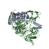
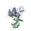
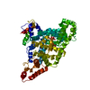
 HOMO SAPIENS (human)
 X-RAY DIFFRACTION /
  Authors
 Citation
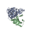
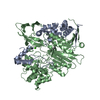
 Structure visualization
 Downloads & links
 Other downloads


 PDBj
PDBj


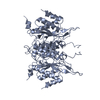
 Assembly
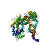

 SPODOPTERA FRUGIPERDA (fall armyworm) / References: UniProt: Q15833
 Mass: 35.453 Da / Num. of mol.: 1 / Source method: obtained synthetically / Formula: Cl
 Mass: 18.015 Da / Num. of mol.: 13 / Source method: isolated from a natural source / Formula: H2O
 Sample preparation
 / Beamline: I03 / Wavelength: 0.976
  Processing