 Movie
Movie Controller


+ Open data

- Basic information
| Entry | Database: PDB / ID: 4c9o | ||||||
|---|---|---|---|---|---|---|---|
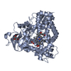
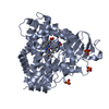
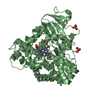
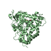
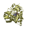
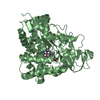
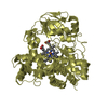
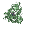
| Title | Structure of Cyanide and Camphor bound D259N mutant of CYP101D1 | ||||||
 Components Components | CYTOCHROME P450 | ||||||
 Keywords Keywords | OXIDOREDUCTASE / MONO-OXYGENASE / P450 | ||||||
| Function / homology |  Function and homology information Function and homology informationoxidoreductase activity, acting on paired donors, with incorporation or reduction of molecular oxygen / monooxygenase activity / iron ion binding / heme binding Similarity search - Function | ||||||
| Biological species |  NOVOSPHINGOBIUM AROMATICIVORANS (bacteria) NOVOSPHINGOBIUM AROMATICIVORANS (bacteria) | ||||||
| Method |  X-RAY DIFFRACTION / SYNCHROTRON / MOLECULAR REPLACEMENT / Resolution: 1.977 Å X-RAY DIFFRACTION / SYNCHROTRON / MOLECULAR REPLACEMENT / Resolution: 1.977 Å | ||||||
 Authors Authors | Batabyal, D. / L Poulos, T. | ||||||
 Citation Citation | Journal: Biochemistry / Year: 2013 Title: Crystal Structures and Functional Characterization of Wild-Type Cyp101D1 and its Active Site Mutants. Authors: Batabyal, D. / Poulos, T.L. | ||||||
| History |
|
- Structure visualization
| Structure viewer | Molecule: MolmilJmol/JSmol |
|---|
- Downloads & links
-Download
| PDBx/mmCIF format | 4c9o.cif.gz | 486.5 KB | Display | PDBx/mmCIF format |
|---|---|---|---|---|
| PDB format | pdb4c9o.ent.gz | 408.1 KB | Display | PDB format |
| PDBx/mmJSON format | 4c9o.json.gz | Tree view | PDBx/mmJSON format | |
| Others |  Other downloads Other downloads |
-Validation report
| Arichive directory | https://data.pdbj.org/pub/pdb/validation_reports/c9/4c9oftp://data.pdbj.org/pub/pdb/validation_reports/c9/4c9o | HTTPS FTP |
|---|
-Related structure data
| Related structure data |  4c9kC  4c9lC  4c9mC  4c9nC  4c9pC  3lxiS C: citing same article ( S: Starting model for refinement |
|---|---|
| Similar structure data |
-Links
 PDBj
PDBj


- Assembly
| Deposited unit | 
| |||||||||
|---|---|---|---|---|---|---|---|---|---|---|
| 1 | 
| |||||||||
| 2 | 
| |||||||||
| Unit cell |
| |||||||||
| Components on special symmetry positions |
|
-Components
| #1: Protein | Mass: 47336.988 Da / Num. of mol.: 2 / Mutation: YES Source method: isolated from a genetically manipulated source Source: (gene. exp.) NOVOSPHINGOBIUM AROMATICIVORANS (bacteria)Strain: DSM12444 / Production host: #2: Chemical |   Mass: 616.487 Da / Num. of mol.: 2 / Source method: obtained synthetically / Formula: C34H32FeN4O4 Mass: 616.487 Da / Num. of mol.: 2 / Source method: obtained synthetically / Formula: C34H32FeN4O4#3: Chemical |   Mass: 152.233 Da / Num. of mol.: 2 / Source method: obtained synthetically / Formula: C10H16O Mass: 152.233 Da / Num. of mol.: 2 / Source method: obtained synthetically / Formula: C10H16O#4: Chemical |   Mass: 26.017 Da / Num. of mol.: 2 / Source method: obtained synthetically / Formula: CN Mass: 26.017 Da / Num. of mol.: 2 / Source method: obtained synthetically / Formula: CN#5: Water | ChemComp-HOH / |  Mass: 18.015 Da / Num. of mol.: 1010 / Source method: isolated from a natural source / Formula: H2O Mass: 18.015 Da / Num. of mol.: 1010 / Source method: isolated from a natural source / Formula: H2O |
|---|
-Experimental details
-Experiment
| Experiment | Method: X-RAY DIFFRACTION / Number of used crystals: 1 |
|---|
- Sample preparation
| Crystal | Density Matthews: 3.49 Å3/Da / Density % sol: 64.8 % / Description: NONE |
|---|---|
| Crystal grow | pH: 8.2 / Details: 100MM TRIS, PH 8.2, 1.6M AMMONIUM SULPHATE |
-Data collection
| Diffraction | Mean temperature: 100 K |
|---|---|
| Diffraction source | Source: SYNCHROTRON / Site: SSRL  / Beamline: BL7-1 / Wavelength: 1 / Beamline: BL7-1 / Wavelength: 1 |
| Detector | Detector: CCD / Date: Mar 30, 2013 |
| Radiation | Protocol: SINGLE WAVELENGTH / Monochromatic (M) / Laue (L): M / Scattering type: x-ray |
| Radiation wavelength | Wavelength: 1 Å / Relative weight: 1 |
| Reflection | Resolution: 1.98→50 Å / Num. obs: 94411 / % possible obs: 99.9 % / Observed criterion σ(I): 5.1 / Redundancy: 14.5 % / Biso Wilson estimate: 23.85 Å2 / Rmerge(I) obs: 0.1 / Net I/σ(I): 24 |
| Reflection shell | Resolution: 1.98→2.09 Å / Redundancy: 14.4 % / Rmerge(I) obs: 0.61 / Mean I/σ(I) obs: 5.1 / % possible all: 99.2 |
- Processing
| Software |
| |||||||||||||||||||||||||||||||||||||||||||||||||||||||||||||||||||||||||||||||||||||||||||||||||||||||||||||||||||||||||||||||||||||||||||||||||||||||||||||||||||||||||||||||||||||||||||||||||||||||||||||||||||||||||||||||||||||||||||||||||||||||||||||||||||||||||||||||||||
|---|---|---|---|---|---|---|---|---|---|---|---|---|---|---|---|---|---|---|---|---|---|---|---|---|---|---|---|---|---|---|---|---|---|---|---|---|---|---|---|---|---|---|---|---|---|---|---|---|---|---|---|---|---|---|---|---|---|---|---|---|---|---|---|---|---|---|---|---|---|---|---|---|---|---|---|---|---|---|---|---|---|---|---|---|---|---|---|---|---|---|---|---|---|---|---|---|---|---|---|---|---|---|---|---|---|---|---|---|---|---|---|---|---|---|---|---|---|---|---|---|---|---|---|---|---|---|---|---|---|---|---|---|---|---|---|---|---|---|---|---|---|---|---|---|---|---|---|---|---|---|---|---|---|---|---|---|---|---|---|---|---|---|---|---|---|---|---|---|---|---|---|---|---|---|---|---|---|---|---|---|---|---|---|---|---|---|---|---|---|---|---|---|---|---|---|---|---|---|---|---|---|---|---|---|---|---|---|---|---|---|---|---|---|---|---|---|---|---|---|---|---|---|---|---|---|---|---|---|---|---|---|---|---|---|---|---|---|---|---|---|---|---|---|---|---|---|---|---|---|---|---|---|---|---|---|---|---|---|---|---|---|---|---|---|---|---|---|---|---|---|---|---|---|---|---|---|
| Refinement | Method to determine structure: MOLECULAR REPLACEMENT Starting model: PDB ENTRY 3LXI Resolution: 1.977→49.802 Å / SU ML: 0.15 / σ(F): 1.34 / Phase error: 15.93 / Stereochemistry target values: ML
| |||||||||||||||||||||||||||||||||||||||||||||||||||||||||||||||||||||||||||||||||||||||||||||||||||||||||||||||||||||||||||||||||||||||||||||||||||||||||||||||||||||||||||||||||||||||||||||||||||||||||||||||||||||||||||||||||||||||||||||||||||||||||||||||||||||||||||||||||||
| Solvent computation | Shrinkage radii: 0.9 Å / VDW probe radii: 1.11 Å / Solvent model: FLAT BULK SOLVENT MODEL | |||||||||||||||||||||||||||||||||||||||||||||||||||||||||||||||||||||||||||||||||||||||||||||||||||||||||||||||||||||||||||||||||||||||||||||||||||||||||||||||||||||||||||||||||||||||||||||||||||||||||||||||||||||||||||||||||||||||||||||||||||||||||||||||||||||||||||||||||||
| Refinement step | Cycle: LAST / Resolution: 1.977→49.802 Å
| |||||||||||||||||||||||||||||||||||||||||||||||||||||||||||||||||||||||||||||||||||||||||||||||||||||||||||||||||||||||||||||||||||||||||||||||||||||||||||||||||||||||||||||||||||||||||||||||||||||||||||||||||||||||||||||||||||||||||||||||||||||||||||||||||||||||||||||||||||
| Refine LS restraints |
| |||||||||||||||||||||||||||||||||||||||||||||||||||||||||||||||||||||||||||||||||||||||||||||||||||||||||||||||||||||||||||||||||||||||||||||||||||||||||||||||||||||||||||||||||||||||||||||||||||||||||||||||||||||||||||||||||||||||||||||||||||||||||||||||||||||||||||||||||||
| LS refinement shell |
| |||||||||||||||||||||||||||||||||||||||||||||||||||||||||||||||||||||||||||||||||||||||||||||||||||||||||||||||||||||||||||||||||||||||||||||||||||||||||||||||||||||||||||||||||||||||||||||||||||||||||||||||||||||||||||||||||||||||||||||||||||||||||||||||||||||||||||||||||||
| Refinement TLS params. | Method: refined / Refine-ID: X-RAY DIFFRACTION
| |||||||||||||||||||||||||||||||||||||||||||||||||||||||||||||||||||||||||||||||||||||||||||||||||||||||||||||||||||||||||||||||||||||||||||||||||||||||||||||||||||||||||||||||||||||||||||||||||||||||||||||||||||||||||||||||||||||||||||||||||||||||||||||||||||||||||||||||||||
| Refinement TLS group |
|
