 Movie
Movie Controller

[English] 日本語
 Yorodumi
- PDB-4c5b: The X-ray crystal structure of D-alanyl-D-alanine ligase in compl... -
+ Open data

- Basic information
| Entry | Database: PDB / ID: 4c5b | ||||||
|---|---|---|---|---|---|---|---|
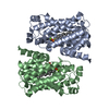
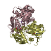
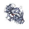
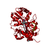
| Title | The X-ray crystal structure of D-alanyl-D-alanine ligase in complex with ATP and D-ala-D-ala | ||||||
 Components Components | D-ALANINE--D-ALANINE LIGASE | ||||||
 Keywords Keywords | LIGASE / DDLB | ||||||
| Function / homology |  Function and homology information Function and homology informationD-alanine-D-alanine ligase / D-alanine-D-alanine ligase activity / peptidoglycan biosynthetic process / cell wall organization / regulation of cell shape / protein homodimerization activity / ATP binding / metal ion binding / cytosol Similarity search - Function | ||||||
| Biological species |  | ||||||
| Method |  X-RAY DIFFRACTION / SYNCHROTRON / MOLECULAR REPLACEMENT / Resolution: 1.5 Å X-RAY DIFFRACTION / SYNCHROTRON / MOLECULAR REPLACEMENT / Resolution: 1.5 Å | ||||||
 Authors Authors | Batson, S. / Majce, V. / Lloyd, A.J. / Rea, D. / Fishwick, C.W.G. / Simmons, K.J. / Fulop, V. / Roper, D.I. | ||||||
 Citation Citation | Journal: Nat Commun / Year: 2017 Title: Inhibition of D-Ala:D-Ala ligase through a phosphorylated form of the antibiotic D-cycloserine. Authors: Batson, S. / de Chiara, C. / Majce, V. / Lloyd, A.J. / Gobec, S. / Rea, D. / Fulop, V. / Thoroughgood, C.W. / Simmons, K.J. / Dowson, C.G. / Fishwick, C.W.G. / de Carvalho, L.P.S. / Roper, D.I. | ||||||
| History |
|
- Structure visualization
| Structure viewer | Molecule: MolmilJmol/JSmol |
|---|
- Downloads & links
-Download
| PDBx/mmCIF format | 4c5b.cif.gz | 260.1 KB | Display | PDBx/mmCIF format |
|---|---|---|---|---|
| PDB format | pdb4c5b.ent.gz | 206.9 KB | Display | PDB format |
| PDBx/mmJSON format | 4c5b.json.gz | Tree view | PDBx/mmJSON format | |
| Others |  Other downloads Other downloads |
-Validation report
| Arichive directory | https://data.pdbj.org/pub/pdb/validation_reports/c5/4c5bftp://data.pdbj.org/pub/pdb/validation_reports/c5/4c5b | HTTPS FTP |
|---|
-Related structure data
| Related structure data |  4c5aC  4c5cC  1iowS C: citing same article ( S: Starting model for refinement |
|---|---|
| Similar structure data |


-Links
 PDBj
PDBj


- Assembly
| Deposited unit | 
| ||||||||
|---|---|---|---|---|---|---|---|---|---|
| 1 |
| ||||||||
| Unit cell |
|
-Components
-Protein , 1 types, 2 molecules AB
| #1: Protein | Mass: 35840.785 Da / Num. of mol.: 2 Source method: isolated from a genetically manipulated source Source: (gene. exp.) References: UniProt: C4ZRI7, UniProt: P07862*PLUS, D-alanine-D-alanine ligase |
|---|
-Non-polymers , 7 types, 783 molecules 


| #2: Chemical |  Mass: 427.201 Da / Num. of mol.: 2 / Source method: obtained synthetically / Formula: C10H15N5O10P2 / Comment: ADP, energy-carrying molecule*YM Mass: 427.201 Da / Num. of mol.: 2 / Source method: obtained synthetically / Formula: C10H15N5O10P2 / Comment: ADP, energy-carrying molecule*YM#3: Chemical | ChemComp-DAL /  Type: D-peptide linking / Mass: 89.093 Da / Num. of mol.: 4 / Source method: obtained synthetically / Formula: C3H7NO2 Type: D-peptide linking / Mass: 89.093 Da / Num. of mol.: 4 / Source method: obtained synthetically / Formula: C3H7NO2#4: Chemical |  Mass: 60.009 Da / Num. of mol.: 2 / Source method: obtained synthetically / Formula: CO3 Mass: 60.009 Da / Num. of mol.: 2 / Source method: obtained synthetically / Formula: CO3#5: Chemical | ChemComp-MG /  Mass: 24.305 Da / Num. of mol.: 6 / Source method: obtained synthetically / Formula: Mg Mass: 24.305 Da / Num. of mol.: 6 / Source method: obtained synthetically / Formula: Mg#6: Chemical |  Mass: 92.094 Da / Num. of mol.: 2 / Source method: obtained synthetically / Formula: C3H8O3 Mass: 92.094 Da / Num. of mol.: 2 / Source method: obtained synthetically / Formula: C3H8O3#7: Chemical | ChemComp-IMD / |  Mass: 69.085 Da / Num. of mol.: 1 / Source method: obtained synthetically / Formula: C3H5N2 Mass: 69.085 Da / Num. of mol.: 1 / Source method: obtained synthetically / Formula: C3H5N2#8: Water | ChemComp-HOH / | Mass: 18.015 Da / Num. of mol.: 766 / Source method: isolated from a natural source / Formula: H2O |
|---|
-Experimental details
-Experiment
| Experiment | Method: X-RAY DIFFRACTION / Number of used crystals: 1 |
|---|
- Sample preparation
| Crystal | Density Matthews: 1.9 Å3/Da / Density % sol: 36 % / Description: NONE |
|---|
-Data collection
| Diffraction | Mean temperature: 100 K |
|---|---|
| Diffraction source | Source: SYNCHROTRON / Site: Diamond  / Beamline: I04 / Wavelength: 0.9763 / Beamline: I04 / Wavelength: 0.9763 |
| Detector | Type: ADSC CCD / Detector: CCD |
| Radiation | Protocol: SINGLE WAVELENGTH / Monochromatic (M) / Laue (L): M / Scattering type: x-ray |
| Radiation wavelength | Wavelength: 0.9763 Å / Relative weight: 1 |
| Reflection | Resolution: 1.5→48 Å / Num. obs: 91443 / % possible obs: 99.9 % / Observed criterion σ(I): 2.1 / Redundancy: 6.6 % / Rmerge(I) obs: 0.09 / Net I/σ(I): 18.9 |
| Reflection shell | Resolution: 1.5→1.55 Å / Rmerge(I) obs: 0.58 / Mean I/σ(I) obs: 2.1 / % possible all: 99.1 |
- Processing
| Software |
| ||||||||||||||||||||||||||||||||||||||||||||||||||||||||||||||||||||||||||||||||||||||||||||||||||||||||||||||||||||||||||||||||||||||||||||||||||||||||||||||||||||||||||||||||||||||
|---|---|---|---|---|---|---|---|---|---|---|---|---|---|---|---|---|---|---|---|---|---|---|---|---|---|---|---|---|---|---|---|---|---|---|---|---|---|---|---|---|---|---|---|---|---|---|---|---|---|---|---|---|---|---|---|---|---|---|---|---|---|---|---|---|---|---|---|---|---|---|---|---|---|---|---|---|---|---|---|---|---|---|---|---|---|---|---|---|---|---|---|---|---|---|---|---|---|---|---|---|---|---|---|---|---|---|---|---|---|---|---|---|---|---|---|---|---|---|---|---|---|---|---|---|---|---|---|---|---|---|---|---|---|---|---|---|---|---|---|---|---|---|---|---|---|---|---|---|---|---|---|---|---|---|---|---|---|---|---|---|---|---|---|---|---|---|---|---|---|---|---|---|---|---|---|---|---|---|---|---|---|---|---|
| Refinement | Method to determine structure: MOLECULAR REPLACEMENT Starting model: PDB ENTRY 1IOW Resolution: 1.5→47.99 Å / Cor.coef. Fo:Fc: 0.966 / Cor.coef. Fo:Fc free: 0.95 / SU B: 2.445 / SU ML: 0.046 / Cross valid method: THROUGHOUT / ESU R: 0.075 / ESU R Free: 0.076 / Stereochemistry target values: MAXIMUM LIKELIHOOD Details: HYDROGENS HAVE BEEN ADDED IN THE RIDING POSITIONS. U VALUES WITH TLS ADDED
| ||||||||||||||||||||||||||||||||||||||||||||||||||||||||||||||||||||||||||||||||||||||||||||||||||||||||||||||||||||||||||||||||||||||||||||||||||||||||||||||||||||||||||||||||||||||
| Solvent computation | Ion probe radii: 0.8 Å / Shrinkage radii: 0.8 Å / VDW probe radii: 1.4 Å / Solvent model: MASK | ||||||||||||||||||||||||||||||||||||||||||||||||||||||||||||||||||||||||||||||||||||||||||||||||||||||||||||||||||||||||||||||||||||||||||||||||||||||||||||||||||||||||||||||||||||||
| Displacement parameters | Biso mean: 16.627 Å2
| ||||||||||||||||||||||||||||||||||||||||||||||||||||||||||||||||||||||||||||||||||||||||||||||||||||||||||||||||||||||||||||||||||||||||||||||||||||||||||||||||||||||||||||||||||||||
| Refinement step | Cycle: LAST / Resolution: 1.5→47.99 Å
| ||||||||||||||||||||||||||||||||||||||||||||||||||||||||||||||||||||||||||||||||||||||||||||||||||||||||||||||||||||||||||||||||||||||||||||||||||||||||||||||||||||||||||||||||||||||
| Refine LS restraints |
|