 Movie
Movie Controller

[English] 日本語
 Yorodumi
- PDB-3h6t: Crystal structure of the iGluR2 ligand-binding core (S1S2J-N754S)... -
+ Open data

- Basic information
| Entry | Database: PDB / ID: 3h6t | ||||||
|---|---|---|---|---|---|---|---|
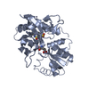
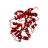
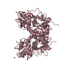
| Title | Crystal structure of the iGluR2 ligand-binding core (S1S2J-N754S) in complex with glutamate and cyclothiazide at 2.25 A resolution | ||||||
 Components Components | Glutamate receptor 2 | ||||||
 Keywords Keywords | MEMBRANE PROTEIN / AMPA receptor ligand-binding core / iGluR2 S1S2J-N754S / cyclothiazide / allosteric modulation | ||||||
| Function / homology |  Function and homology information Function and homology informationspine synapse / dendritic spine neck / dendritic spine cytoplasm / dendritic spine head / cellular response to amine stimulus / Activation of AMPA receptors / ligand-gated monoatomic cation channel activity / perisynaptic space / Trafficking of GluR2-containing AMPA receptors / response to lithium ion ...spine synapse / dendritic spine neck / dendritic spine cytoplasm / dendritic spine head / cellular response to amine stimulus / Activation of AMPA receptors / ligand-gated monoatomic cation channel activity / perisynaptic space / Trafficking of GluR2-containing AMPA receptors / response to lithium ion / AMPA glutamate receptor activity / AMPA glutamate receptor clustering / kainate selective glutamate receptor activity / immunoglobulin binding / AMPA glutamate receptor complex / regulation of receptor recycling / extracellularly glutamate-gated ion channel activity / cellular response to glycine / ionotropic glutamate receptor complex / asymmetric synapse / Unblocking of NMDA receptors, glutamate binding and activation / glutamate receptor binding / positive regulation of synaptic transmission / conditioned place preference / response to fungicide / regulation of synaptic transmission, glutamatergic / extracellular ligand-gated monoatomic ion channel activity / cytoskeletal protein binding / glutamate-gated receptor activity / cellular response to brain-derived neurotrophic factor stimulus / regulation of long-term synaptic depression / somatodendritic compartment / glutamate-gated calcium ion channel activity / presynaptic active zone membrane / excitatory synapse / ionotropic glutamate receptor signaling pathway / ionotropic glutamate receptor binding / dendrite cytoplasm / dendrite membrane / ligand-gated monoatomic ion channel activity involved in regulation of presynaptic membrane potential / positive regulation of excitatory postsynaptic potential / dendritic shaft / SNARE binding / synaptic membrane / PDZ domain binding / establishment of protein localization / protein tetramerization / synaptic transmission, glutamatergic / transmitter-gated monoatomic ion channel activity involved in regulation of postsynaptic membrane potential / cerebral cortex development / receptor internalization / postsynaptic density membrane / modulation of chemical synaptic transmission / Schaffer collateral - CA1 synapse / terminal bouton / synaptic vesicle / long-term synaptic potentiation / amyloid-beta binding / synaptic vesicle membrane / growth cone / presynapse / signaling receptor activity / presynaptic membrane / scaffold protein binding / dendritic spine / chemical synaptic transmission / perikaryon / postsynaptic membrane / neuron projection / postsynaptic density / external side of plasma membrane / axon / neuronal cell body / synapse / dendrite / protein kinase binding / protein-containing complex binding / glutamatergic synapse / cell surface / endoplasmic reticulum / protein-containing complex / membrane / identical protein binding / plasma membrane Similarity search - Function | ||||||
| Biological species |  | ||||||
| Method |  X-RAY DIFFRACTION / SYNCHROTRON / MOLECULAR REPLACEMENT / molecular replacement / Resolution: 2.25 Å X-RAY DIFFRACTION / SYNCHROTRON / MOLECULAR REPLACEMENT / molecular replacement / Resolution: 2.25 Å | ||||||
 Authors Authors | Hald, H. / Gajhede, M. / Kastrup, J.S. | ||||||
 Citation Citation | Journal: J.Mol.Biol. / Year: 2009 Title: Distinct structural features of cyclothiazide are responsible for effects on peak current amplitude and desensitization kinetics at iGluR2. Authors: Hald, H. / Ahring, P.K. / Timmermann, D.B. / Liljefors, T. / Gajhede, M. / Kastrup, J.S. #1: Journal: Neuron / Year: 2000Title: Mechanisms for activation and antagonism of an AMPA-sensitive glutamate receptor: crystal structures of the GluR2 ligand binding core. Authors: Armstrong, N. / Gouaux, E. #2: Journal: Nature / Year: 2002Title: Mechanism of glutamate receptor desensitization. Authors: Sun, Y. / Olson, R. / Horning, M. / Armstrong, N. / Mayer, M. / Gouaux, E. | ||||||
| History |
|
- Structure visualization
| Structure viewer | Molecule: MolmilJmol/JSmol |
|---|
- Downloads & links
-Download
| PDBx/mmCIF format | 3h6t.cif.gz | 197.1 KB | Display | PDBx/mmCIF format |
|---|---|---|---|---|
| PDB format | pdb3h6t.ent.gz | 153.8 KB | Display | PDB format |
| PDBx/mmJSON format | 3h6t.json.gz | Tree view | PDBx/mmJSON format | |
| Others |  Other downloads Other downloads |
-Validation report
| Arichive directory | https://data.pdbj.org/pub/pdb/validation_reports/h6/3h6tftp://data.pdbj.org/pub/pdb/validation_reports/h6/3h6t | HTTPS FTP |
|---|
-Related structure data
| Related structure data | 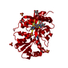 3h6uC 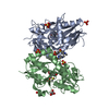 3h6vC 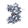 3h6wC  1ftjS C: citing same article ( S: Starting model for refinement |
|---|---|
| Similar structure data |


-Links
 PDBj
PDBj


- Assembly
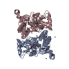
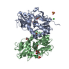
| Deposited unit | 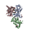
| ||||||||
|---|---|---|---|---|---|---|---|---|---|
| 1 | 
| ||||||||
| 2 | 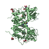
| ||||||||
| Unit cell |
|
-Components
-Protein , 1 types, 3 molecules ABC
| #1: Protein | Mass: 29194.658 Da / Num. of mol.: 3 Fragment: iGluR2-flop ligand-binding core: UNP residues 413-796 Mutation: N754S Source method: isolated from a genetically manipulated source Source: (gene. exp.)  |
|---|
-Non-polymers , 8 types, 1060 molecules 


| #2: Chemical |  Type: L-peptide linking / Mass: 147.129 Da / Num. of mol.: 3 / Source method: obtained synthetically / Formula: C5H9NO4 Type: L-peptide linking / Mass: 147.129 Da / Num. of mol.: 3 / Source method: obtained synthetically / Formula: C5H9NO4#3: Chemical |  Mass: 389.878 Da / Num. of mol.: 3 / Source method: obtained synthetically / Formula: C14H16ClN3O4S2 Mass: 389.878 Da / Num. of mol.: 3 / Source method: obtained synthetically / Formula: C14H16ClN3O4S2#4: Chemical | ChemComp-ZN /  Mass: 65.409 Da / Num. of mol.: 10 / Source method: obtained synthetically / Formula: Zn Mass: 65.409 Da / Num. of mol.: 10 / Source method: obtained synthetically / Formula: Zn#5: Chemical | ChemComp-GOL /  Mass: 92.094 Da / Num. of mol.: 9 / Source method: obtained synthetically / Formula: C3H8O3 Mass: 92.094 Da / Num. of mol.: 9 / Source method: obtained synthetically / Formula: C3H8O3#6: Chemical |  Mass: 59.044 Da / Num. of mol.: 2 / Source method: obtained synthetically / Formula: C2H3O2 Mass: 59.044 Da / Num. of mol.: 2 / Source method: obtained synthetically / Formula: C2H3O2#7: Chemical | ChemComp-DMS / |  Mass: 78.133 Da / Num. of mol.: 1 / Source method: obtained synthetically / Formula: C2H6OS / Comment: DMSO, precipitant*YM Mass: 78.133 Da / Num. of mol.: 1 / Source method: obtained synthetically / Formula: C2H6OS / Comment: DMSO, precipitant*YM#8: Chemical | ChemComp-CAC / |  Mass: 136.989 Da / Num. of mol.: 1 / Source method: obtained synthetically / Formula: C2H6AsO2 Mass: 136.989 Da / Num. of mol.: 1 / Source method: obtained synthetically / Formula: C2H6AsO2#9: Water | ChemComp-HOH / | Mass: 18.015 Da / Num. of mol.: 1031 / Source method: isolated from a natural source / Formula: H2O |
|---|
-Details
| Has protein modification | Y |
|---|---|
| Sequence details | NATIVE IGLUR2 IS A MEMBRANE PROTEIN. THE PROTEIN CRYSTALLIZED IS THE EXTRACELLULAR LIGAND-BINDING ...NATIVE IGLUR2 IS A MEMBRANE PROTEIN. THE PROTEIN CRYSTALLIZ |
-Experimental details
-Experiment
| Experiment | Method: X-RAY DIFFRACTION / Number of used crystals: 1 |
|---|
- Sample preparation
| Crystal | Density Matthews: 2.52 Å3/Da / Density % sol: 51.28 % |
|---|---|
| Crystal grow | Temperature: 279 K / pH: 6.5 Details: 25 % PEG 4000, 0.3 M Zinc acetate, 0.1 M Cacodylate pH 6.5, VAPOR DIFFUSION, HANGING DROP, temperature 279K |
-Data collection
| Diffraction | Mean temperature: 96.5 K |
|---|---|
| Diffraction source | Source: SYNCHROTRON / Site: MAX II  / Beamline: I711 / Wavelength: 1.141 / Beamline: I711 / Wavelength: 1.141 |
| Detector | Type: MAR CCD 165 mm / Detector: CCD / Date: Dec 14, 2005 |
| Radiation | Monochromator: SINGLE ASYMMETRICALLY CUT SI(111) CRYSTAL WITH HORIZONTAL DIFFRACTION PLANE. THE CRYSTAL IS BENDABLE FOR HORIZONTAL FOCUSING. Protocol: SINGLE WAVELENGTH / Monochromatic (M) / Laue (L): M / Scattering type: x-ray |
| Radiation wavelength | Wavelength: 1.141 Å / Relative weight: 1 |
| Reflection | Resolution: 2.25→22.84 Å / Num. obs: 42613 / % possible obs: 99.3 % / Redundancy: 3.4 % / Biso Wilson estimate: 31.1 Å2 / Rmerge(I) obs: 0.084 / Rsym value: 0.084 / Net I/σ(I): 7.8 |
| Reflection shell | Resolution: 2.25→2.37 Å / Redundancy: 3.4 % / Rmerge(I) obs: 0.346 / Mean I/σ(I) obs: 1.9 / Rsym value: 0.346 / % possible all: 100 |
-Phasing
| Phasing | Method: molecular replacement | |||||||||
|---|---|---|---|---|---|---|---|---|---|---|
| Phasing MR | Model details: Phaser MODE: MR_AUTO
|
- Processing
| Software |
| ||||||||||||||||||||||||||||||||||||||||||||||||||||||||||||||||||||||||||||||||
|---|---|---|---|---|---|---|---|---|---|---|---|---|---|---|---|---|---|---|---|---|---|---|---|---|---|---|---|---|---|---|---|---|---|---|---|---|---|---|---|---|---|---|---|---|---|---|---|---|---|---|---|---|---|---|---|---|---|---|---|---|---|---|---|---|---|---|---|---|---|---|---|---|---|---|---|---|---|---|---|---|---|
| Refinement | Method to determine structure: MOLECULAR REPLACEMENT Starting model: PDB ENTRY 1FTJ Resolution: 2.25→22.84 Å / Rfactor Rfree error: 0.005 / Occupancy max: 1 / Occupancy min: 0.5 / Data cutoff high absF: 2075455.12 / Data cutoff low absF: 0 / Isotropic thermal model: RESTRAINED / Cross valid method: THROUGHOUT / σ(F): 0 / Stereochemistry target values: ENGH & HUBER
| ||||||||||||||||||||||||||||||||||||||||||||||||||||||||||||||||||||||||||||||||
| Solvent computation | Solvent model: FLAT MODEL / Bsol: 46.16 Å2 / ksol: 0.315591 e/Å3 | ||||||||||||||||||||||||||||||||||||||||||||||||||||||||||||||||||||||||||||||||
| Displacement parameters | Biso mean: 29.1 Å2
| ||||||||||||||||||||||||||||||||||||||||||||||||||||||||||||||||||||||||||||||||
| Refine analyze |
| ||||||||||||||||||||||||||||||||||||||||||||||||||||||||||||||||||||||||||||||||
| Refinement step | Cycle: LAST / Resolution: 2.25→22.84 Å
| ||||||||||||||||||||||||||||||||||||||||||||||||||||||||||||||||||||||||||||||||
| Refine LS restraints |
| ||||||||||||||||||||||||||||||||||||||||||||||||||||||||||||||||||||||||||||||||
| LS refinement shell | Resolution: 2.25→2.27 Å / Rfactor Rfree error: 0.054 / Total num. of bins used: 41
| ||||||||||||||||||||||||||||||||||||||||||||||||||||||||||||||||||||||||||||||||
| Xplor file |
|
