[English] 日本語
Yorodumi
- PDB-6giv: Structure of GluA2-N775S ligand-binding domain (S1S2J) in complex... -

+
Open data


ID or keywords:

Loading...

-
Basic information

Entry
Database: PDB / ID: 6giv
TitleStructure of GluA2-N775S ligand-binding domain (S1S2J) in complex with glutamate and Rubidium Bromide at 1.75 A resolution
ComponentsGlutamate receptor 2
KeywordsMEMBRANE PROTEIN / Ionotropic glutamate receptor / GluA2-N775S ligand-binding domain / agonist / bromide ions
Function / homology
Function and homology information


spine synapse / dendritic spine neck / dendritic spine head / Activation of AMPA receptors / perisynaptic space / AMPA glutamate receptor activity / Trafficking of GluR2-containing AMPA receptors / response to lithium ion / immunoglobulin binding / AMPA glutamate receptor complex ...spine synapse / dendritic spine neck / dendritic spine head / Activation of AMPA receptors / perisynaptic space / AMPA glutamate receptor activity / Trafficking of GluR2-containing AMPA receptors / response to lithium ion / immunoglobulin binding / AMPA glutamate receptor complex / kainate selective glutamate receptor activity / ionotropic glutamate receptor complex / extracellularly glutamate-gated ion channel activity / cellular response to glycine / asymmetric synapse / regulation of receptor recycling / Unblocking of NMDA receptors, glutamate binding and activation / glutamate receptor binding / positive regulation of synaptic transmission / glutamate-gated receptor activity / presynaptic active zone membrane / response to fungicide / regulation of synaptic transmission, glutamatergic / somatodendritic compartment / cellular response to brain-derived neurotrophic factor stimulus / dendrite membrane / ligand-gated monoatomic ion channel activity involved in regulation of presynaptic membrane potential / cytoskeletal protein binding / ionotropic glutamate receptor signaling pathway / dendrite cytoplasm / SNARE binding / dendritic shaft / synaptic membrane / synaptic transmission, glutamatergic / transmitter-gated monoatomic ion channel activity involved in regulation of postsynaptic membrane potential / PDZ domain binding / protein tetramerization / postsynaptic density membrane / ionotropic glutamate receptor binding / Schaffer collateral - CA1 synapse / modulation of chemical synaptic transmission / establishment of protein localization / terminal bouton / receptor internalization / cerebral cortex development / synaptic vesicle membrane / synaptic vesicle / presynapse / presynaptic membrane / signaling receptor activity / amyloid-beta binding / growth cone / scaffold protein binding / chemical synaptic transmission / postsynaptic membrane / perikaryon / dendritic spine / postsynaptic density / neuron projection / axon / neuronal cell body / glutamatergic synapse / synapse / dendrite / protein-containing complex binding / endoplasmic reticulum membrane / protein kinase binding / cell surface / endoplasmic reticulum / protein-containing complex / identical protein binding / membrane / plasma membrane
Similarity search - Function
Bacterial extracellular solute-binding proteins, family 3 / Solute-binding protein family 3/N-terminal domain of MltF / Ionotropic glutamate receptor, metazoa / Ligated ion channel L-glutamate- and glycine-binding site / : / Ligand-gated ion channel / Ionotropic glutamate receptor, L-glutamate and glycine-binding domain / Ligated ion channel L-glutamate- and glycine-binding site / Ionotropic glutamate receptor / Eukaryotic homologues of bacterial periplasmic substrate binding proteins. ...Bacterial extracellular solute-binding proteins, family 3 / Solute-binding protein family 3/N-terminal domain of MltF / Ionotropic glutamate receptor, metazoa / Ligated ion channel L-glutamate- and glycine-binding site / : / Ligand-gated ion channel / Ionotropic glutamate receptor, L-glutamate and glycine-binding domain / Ligated ion channel L-glutamate- and glycine-binding site / Ionotropic glutamate receptor / Eukaryotic homologues of bacterial periplasmic substrate binding proteins. / Receptor, ligand binding region / Receptor family ligand binding region / Periplasmic binding protein-like II / Periplasmic binding protein-like I / D-Maltodextrin-Binding Protein; domain 2 / 3-Layer(aba) Sandwich / Alpha Beta
Similarity search - Domain/homology
BROMIDE ION / GLUTAMIC ACID / Glutamate receptor 2
Similarity search - Component
Biological speciesRattus norvegicus (Norway rat)
MethodX-RAY DIFFRACTION / SYNCHROTRON / MOLECULAR REPLACEMENT / molecular replacement / Resolution: 1.75 Å
AuthorsVenskutonyte, R. / Frydenvang, K. / Kastrup, J.S.
CitationJournal: Neuron / Year: 2019
Title: Nanoscale Mobility of the Apo State and TARP Stoichiometry Dictate the Gating Behavior of Alternatively Spliced AMPA Receptors.
Authors: Dawe, G.B. / Kadir, M.F. / Venskutonyte, R. / Perozzo, A.M. / Yan, Y. / Alexander, R.P.D. / Navarrete, C. / Santander, E.A. / Arsenault, M. / Fuentes, C. / Aurousseau, M.R.P. / Frydenvang, K. ...Authors: Dawe, G.B. / Kadir, M.F. / Venskutonyte, R. / Perozzo, A.M. / Yan, Y. / Alexander, R.P.D. / Navarrete, C. / Santander, E.A. / Arsenault, M. / Fuentes, C. / Aurousseau, M.R.P. / Frydenvang, K. / Barrera, N.P. / Kastrup, J.S. / Edwardson, J.M. / Bowie, D.
History
DepositionMay 15, 2018Deposition site: PDBE / Processing site: PDBE
Revision 1.0May 15, 2019Provider: repository / Type: Initial release
Revision 1.1Jun 19, 2019Group: Data collection / Database references / Category: citation / pdbx_database_proc / Item: _citation.journal_volume / _citation.page_first
Revision 1.2Jan 17, 2024Group: Data collection / Database references / Refinement description
Category: chem_comp_atom / chem_comp_bond ...chem_comp_atom / chem_comp_bond / database_2 / pdbx_initial_refinement_model
Item: _database_2.pdbx_DOI / _database_2.pdbx_database_accession

-
Structure visualization

Structure viewerMolecule:
MolmilJmol/JSmol

Downloads & links

-
Assembly

Deposited unit
A: Glutamate receptor 2
hetero molecules


Theoretical massNumber of molelcules
Total (without water)29,8517
Polymers29,2521
Non-polymers5996
Water4,756264
1
A: Glutamate receptor 2
hetero molecules

A: Glutamate receptor 2
hetero molecules


Theoretical massNumber of molelcules
Total (without water)59,70214
Polymers58,5032
Non-polymers1,19912
Water362
TypeNameSymmetry operationNumber
identity operation1_555x,y,z1
crystal symmetry operation2_556-x,y,-z+11
Buried area3690 Å2
ΔGint-39 kcal/mol
Surface area24030 Å2
MethodPISA
Unit cell
Length a, b, c (Å)122.956, 47.513, 49.810
Angle α, β, γ (deg.)90.000, 110.160, 90.000
Int Tables number5
Space group name H-MC121
Components on special symmetry positions
IDModelComponents
11A-583-

HOH

-
Components

-
Protein , 1 types, 1 molecules A

#1: Protein Glutamate receptor 2 / GluR-2 / AMPA-selective glutamate receptor 2 / GluR-B / GluR-K2 / Glutamate receptor ionotropic / ...GluR-2 / AMPA-selective glutamate receptor 2 / GluR-B / GluR-K2 / Glutamate receptor ionotropic / AMPA 2 / GluA2


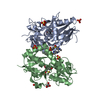
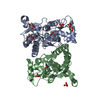
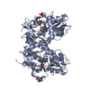
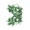
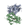
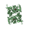
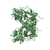
Mass: 29251.709 Da / Num. of mol.: 1 / Mutation: N775S,N775S
Source method: isolated from a genetically manipulated source
Details: The protein crystallized is the extracellular ligand-binding domain of GluA2-N775S. Transmembrane regions were genetically removed and replaced with a Gly-Thr linker (residue 118-119). The ...Details: The protein crystallized is the extracellular ligand-binding domain of GluA2-N775S. Transmembrane regions were genetically removed and replaced with a Gly-Thr linker (residue 118-119). The sequence matches discontinously with the reference database (UNP residues 413-527 and 653-797, numbering with signal peptide of 21 amino acids). The two first residues (GLY, ALA) are cloning remnants.
Source: (gene. exp.) Rattus norvegicus (Norway rat) / Gene: Gria2, Glur2 / Plasmid: pET-22b(+) / Production host: Escherichia coli BL21(DE3) (bacteria) / Variant (production host): Origami B (DE3) / References: UniProt: P19491

-
Non-polymers , 5 types, 270 molecules

#2: Chemical ChemComp-GLU / GLUTAMIC ACID


Type: L-peptide linking / Mass: 147.129 Da / Num. of mol.: 1 / Source method: obtained synthetically / Formula: C5H9NO4
#3: Chemical ChemComp-SO4 / SULFATE ION


Mass: 96.063 Da / Num. of mol.: 1 / Source method: obtained synthetically / Formula: SO4
#4: Chemical ChemComp-GOL / GLYCEROL / GLYCERIN / PROPANE-1,2,3-TRIOL


Mass: 92.094 Da / Num. of mol.: 3 / Source method: obtained synthetically / Formula: C3H8O3
#5: Chemical ChemComp-BR / BROMIDE ION


Mass: 79.904 Da / Num. of mol.: 1 / Source method: obtained synthetically / Formula: Br
#6: Water ChemComp-HOH / water


Mass: 18.015 Da / Num. of mol.: 264 / Source method: isolated from a natural source / Formula: H2O

-
Experimental details

-
Experiment

ExperimentMethod: X-RAY DIFFRACTION / Number of used crystals: 1

-
Sample preparation

CrystalDensity Matthews: 2.33 Å3/Da / Density % sol: 47.31 %
Crystal growTemperature: 279 K / Method: vapor diffusion, hanging drop / pH: 6.5
Details: 18-24.4% PEG4000, 0.2-0.3 M Li2SO4, and 0.1 M cacodylate, pH 6.5

-
Data collection

DiffractionMean temperature: 100 K
Diffraction sourceSource: SYNCHROTRON / Site: MAX II / Beamline: I911-3 / Wavelength: 0.91976 Å
DetectorType: MARMOSAIC 225 mm CCD / Detector: CCD / Date: Sep 30, 2014
RadiationProtocol: SINGLE WAVELENGTH / Monochromatic (M) / Laue (L): M / Scattering type: x-ray
Radiation wavelengthWavelength: 0.91976 Å / Relative weight: 1
ReflectionResolution: 1.75→46.76 Å / Num. obs: 27430 / % possible obs: 100 % / Redundancy: 5 % / Biso Wilson estimate: 15.4 Å2 / Rpim(I) all: 0.038 / Rrim(I) all: 0.089 / Rsym value: 0.074 / Net I/av σ(I): 7.5 / Net I/σ(I): 11.4
Reflection shell

Diffraction-ID: 1

Resolution (Å)Redundancy (%)Rmerge(I) obsMean I/σ(I) obsNum. unique obsRpim(I) allRrim(I) allRsym value% possible all
1.75-1.844.70.3731.839980.2030.4510.373100
1.84-1.9650.262.637400.1360.3140.26100
1.96-2.095.10.1534.435530.0810.1850.153100
2.09-2.265.10.1195.332830.0620.1430.119100
2.26-2.475.10.097.730470.0470.1090.09100
2.47-2.775.10.0719.327700.0360.0850.071100
2.77-3.25.10.05910.524190.030.070.059100
3.2-3.915.10.05311.920820.0260.0620.053100
3.91-5.5350.04413.916260.0210.0510.044100
5.53-46.764.80.0414.99120.020.0470.0499.9

-
Phasing

PhasingMethod: molecular replacement
Phasing MRModel details: Phaser MODE: MR_AUTO
Highest resolutionLowest resolution
Rotation1.75 Å43.94 Å
Translation1.75 Å43.94 Å

-
Processing

Software
NameVersionClassification
SCALA3.3.20data scaling
PHASER2.5.1phasing
PHENIXrefinement
PDB_EXTRACT3.24data extraction
XDSdata reduction
RefinementMethod to determine structure: MOLECULAR REPLACEMENT
Starting model: 4O3A
Resolution: 1.75→34.346 Å / SU ML: 0.17 / Cross valid method: THROUGHOUT / σ(F): 1.35 / Phase error: 19.59
RfactorNum. reflection% reflection
Rfree0.1904 1376 5.02 %
Rwork0.1561 --
obs0.1578 27411 99.94 %
Solvent computationShrinkage radii: 0.9 Å / VDW probe radii: 1.11 Å
Displacement parametersBiso max: 112.1 Å2 / Biso mean: 23.9456 Å2 / Biso min: 9 Å2
Refinement stepCycle: final / Resolution: 1.75→34.346 Å
ProteinNucleic acidLigandSolventTotal
Num. atoms2037 0 34 267 2338
Biso mean--43.36 29.21 -
Num. residues----262
Refine LS restraints
Refine-IDTypeDev idealNumber
X-RAY DIFFRACTIONf_bond_d0.0052158
X-RAY DIFFRACTIONf_angle_d0.7522914
X-RAY DIFFRACTIONf_chiral_restr0.049324
X-RAY DIFFRACTIONf_plane_restr0.005363
X-RAY DIFFRACTIONf_dihedral_angle_d13.2491328
LS refinement shell

Refine-ID: X-RAY DIFFRACTION / Rfactor Rfree error: 0 / Total num. of bins used: 10 / % reflection obs: 100 %

Resolution (Å)Rfactor RfreeNum. reflection RfreeRfactor RworkNum. reflection RworkNum. reflection all
1.75-1.81260.25191280.207526082736
1.8126-1.88510.22951520.18525472699
1.8851-1.97090.28341330.227626072740
1.9709-2.07480.21371310.16125892720
2.0748-2.20480.19361310.146625712702
2.2048-2.3750.22811340.180326162750
2.375-2.61390.17131300.155925962726
2.6139-2.9920.18591340.1526242758
2.992-3.76880.18111460.140626012747
3.7688-34.35270.14771570.131326762833
Refinement TLS params.

Method: refined / Refine-ID: X-RAY DIFFRACTION

IDL112)L122)L132)L222)L232)L332)S11 (Å °)S12 (Å °)S13 (Å °)S21 (Å °)S22 (Å °)S23 (Å °)S31 (Å °)S32 (Å °)S33 (Å °)T112)T122)T132)T222)T232)T332)Origin x (Å)Origin y (Å)Origin z (Å)
12.10140.43340.35142.6785-0.11372.45520.02650.1432-0.1616-0.13370.01360.0292-0.0273-0.0291-0.02270.15070.0034-0.05220.1057-0.0090.1273-22.129412.28254.5892
23.8874-1.3092-2.12672.27791.54193.89920.01510.0886-0.06480.0621-0.02330.06450.1151-0.21130.09140.1008-0.0125-0.05190.0777-0.00070.1337-26.07517.501313.6771
36.7661-0.94971.37887.66661.5161.1661-0.0451-0.6132-0.25270.39860.03080.99620.1894-0.00370.06020.1356-0.00410.02170.12660.01690.2445-29.37394.233924.6411
40.3906-0.5879-0.33532.57880.16650.52860.0001-0.0141-0.01890.03190.0178-0.04380.01590.0242-0.0140.1014-0.0044-0.050.11230.01040.1235-20.156616.437222.6899
51.513-0.3654-0.13031.8517-0.69053.2612-0.0078-0.175-0.0790.08950.0840.1735-0.0803-0.1701-0.0730.10750.0033-0.0310.10970.02280.1303-29.164726.585828.4168
64.6537-3.80794.20545.907-4.95396.95680.08360.03760.0329-0.0308-0.0362-0.11690.09920.05790.01530.1522-0.0173-0.01770.11020.00240.1529-21.364732.168220.8283
72.65460.4018-0.85952.9599-0.94052.2655-0.1198-0.0278-0.2861-0.1370.0211-0.05550.16710.09040.10650.09450.0069-0.05450.1025-0.00540.1187-10.8367.113715.9855
85.0758-3.4922-1.35097.7588-0.2873.98160.15790.30220.4717-0.1039-0.1603-0.2991-0.327-0.1034-0.01930.1944-0.0373-0.05480.14750.04660.1407-13.165925.77888.0679
Refinement TLS group
IDRefine-IDRefine TLS-IDSelection detailsAuth asym-IDAuth seq-ID
1X-RAY DIFFRACTION1chain 'A' and (resid 1 through 47 )A1 - 47
2X-RAY DIFFRACTION2chain 'A' and (resid 48 through 65 )A48 - 65
3X-RAY DIFFRACTION3chain 'A' and (resid 66 through 79 )A66 - 79
4X-RAY DIFFRACTION4chain 'A' and (resid 80 through 123 )A80 - 123
5X-RAY DIFFRACTION5chain 'A' and (resid 124 through 202 )A124 - 202
6X-RAY DIFFRACTION6chain 'A' and (resid 203 through 217 )A203 - 217
7X-RAY DIFFRACTION7chain 'A' and (resid 218 through 243 )A218 - 243
8X-RAY DIFFRACTION8chain 'A' and (resid 244 through 262 )A244 - 262

+
About Yorodumi

-
News

-
Feb 9, 2022. New format data for meta-information of EMDB entries

New format data for meta-information of EMDB entries

  • Version 3 of the EMDB header file is now the official format.
  • The previous official version 1.9 will be removed from the archive.

Related info.:EMDB header

External links:wwPDB to switch to version 3 of the EMDB data model

-
Aug 12, 2020. Covid-19 info

Covid-19 info

URL: https://pdbjlvh1.pdbj.org/emnavi/covid19.php

New page: Covid-19 featured information page in EM Navigator.

Related info.:Covid-19 info / Mar 5, 2020. Novel coronavirus structure data

+
Mar 5, 2020. Novel coronavirus structure data

Novel coronavirus structure data

Related info.:Yorodumi Speices / Aug 12, 2020. Covid-19 info

External links:COVID-19 featured content - PDBj / Molecule of the Month (242):Coronavirus Proteases

+
Jan 31, 2019. EMDB accession codes are about to change! (news from PDBe EMDB page)

EMDB accession codes are about to change! (news from PDBe EMDB page)

  • The allocation of 4 digits for EMDB accession codes will soon come to an end. Whilst these codes will remain in use, new EMDB accession codes will include an additional digit and will expand incrementally as the available range of codes is exhausted. The current 4-digit format prefixed with “EMD-” (i.e. EMD-XXXX) will advance to a 5-digit format (i.e. EMD-XXXXX), and so on. It is currently estimated that the 4-digit codes will be depleted around Spring 2019, at which point the 5-digit format will come into force.
  • The EM Navigator/Yorodumi systems omit the EMD- prefix.

Related info.:Q: What is EMD? / ID/Accession-code notation in Yorodumi/EM Navigator

External links:EMDB Accession Codes are Changing Soon! / Contact to PDBj

+
Jul 12, 2017. Major update of PDB

Major update of PDB

  • wwPDB released updated PDB data conforming to the new PDBx/mmCIF dictionary.
  • This is a major update changing the version number from 4 to 5, and with Remediation, in which all the entries are updated.
  • In this update, many items about electron microscopy experimental information are reorganized (e.g. em_software).
  • Now, EM Navigator and Yorodumi are based on the updated data.

External links:wwPDB Remediation / Enriched Model Files Conforming to OneDep Data Standards Now Available in the PDB FTP Archive

-
Yorodumi

Thousand views of thousand structures

  • Yorodumi is a browser for structure data from EMDB, PDB, SASBDB, etc.
  • This page is also the successor to EM Navigator detail page, and also detail information page/front-end page for Omokage search.
  • The word "yorodu" (or yorozu) is an old Japanese word meaning "ten thousand". "mi" (miru) is to see.

Related info.:EMDB / PDB / SASBDB / Comparison of 3 databanks / Yorodumi Search / Aug 31, 2016. New EM Navigator & Yorodumi / Yorodumi Papers / Jmol/JSmol / Function and homology information / Changes in new EM Navigator and Yorodumi

Read more