 Movie
Movie Controller


+ Open data

- Basic information
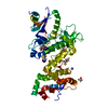
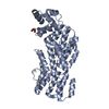
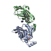
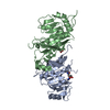
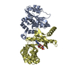
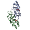
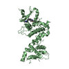
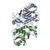
| Entry | Database: PDB / ID: 4c59 | ||||||
|---|---|---|---|---|---|---|---|
| Title | Structure of GAK kinase in complex with nanobody (NbGAK_4) | ||||||
 Components Components |
| ||||||
 Keywords Keywords | TRANSFERASE / KINASE / CONFORMATIONAL PLASTICITY / ACTIVATION | ||||||
| Function / homology |  Function and homology information Function and homology informationregulation of clathrin coat assembly / synaptic vesicle uncoating / Golgi to lysosome transport / clathrin coat disassembly / protein localization to Golgi apparatus / clathrin coat assembly / clathrin-dependent endocytosis / endoplasmic reticulum organization / clathrin-coated vesicle / clathrin binding ...regulation of clathrin coat assembly / synaptic vesicle uncoating / Golgi to lysosome transport / clathrin coat disassembly / protein localization to Golgi apparatus / clathrin coat assembly / clathrin-dependent endocytosis / endoplasmic reticulum organization / clathrin-coated vesicle / clathrin binding / Golgi Associated Vesicle Biogenesis / Golgi organization / intracellular transport / protein folding chaperone / receptor-mediated endocytosis / cyclin binding / protein localization to plasma membrane / negative regulation of neuron projection development / Clathrin-mediated endocytosis / protein folding / presynapse / vesicle / non-specific serine/threonine protein kinase / protein serine kinase activity / focal adhesion / protein serine/threonine kinase activity / perinuclear region of cytoplasm / Golgi apparatus / ATP binding / membrane / cytoplasm / cytosol Similarity search - Function | ||||||
| Biological species |  Homo sapiens (human) Homo sapiens (human) | ||||||
| Method |  X-RAY DIFFRACTION / SYNCHROTRON / MOLECULAR REPLACEMENT / Resolution: 2.8 Å X-RAY DIFFRACTION / SYNCHROTRON / MOLECULAR REPLACEMENT / Resolution: 2.8 Å | ||||||
 Authors Authors | Chaikuad, A. / Keates, T. / Allerston, C.K. / Gileadi, O. / von Delft, F. / Arrowsmith, C.H. / Edwards, A.M. / Bountra, C. / Knapp, S. / Muller-Knapp, S. | ||||||
 Citation Citation | Journal: Biochem. J. / Year: 2014 Title: Structure of cyclin G-associated kinase (GAK) trapped in different conformations using nanobodies. Authors: Chaikuad, A. / Keates, T. / Vincke, C. / Kaufholz, M. / Zenn, M. / Zimmermann, B. / Gutierrez, C. / Zhang, R.G. / Hatzos-Skintges, C. / Joachimiak, A. / Muyldermans, S. / Herberg, F.W. / Knapp, S. / Muller, S. | ||||||
| History |
|
- Structure visualization
| Structure viewer | Molecule: MolmilJmol/JSmol |
|---|
- Downloads & links
-Download
| PDBx/mmCIF format | 4c59.cif.gz | 169.2 KB | Display | PDBx/mmCIF format |
|---|---|---|---|---|
| PDB format | pdb4c59.ent.gz | 133.2 KB | Display | PDB format |
| PDBx/mmJSON format | 4c59.json.gz | Tree view | PDBx/mmJSON format | |
| Others |  Other downloads Other downloads |
-Validation report
| Arichive directory | https://data.pdbj.org/pub/pdb/validation_reports/c5/4c59ftp://data.pdbj.org/pub/pdb/validation_reports/c5/4c59 | HTTPS FTP |
|---|
-Related structure data
| Related structure data | 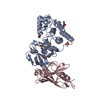 4c57C 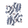 4c58C 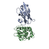 1op9S  3ll6 C: citing same article ( S: Starting model for refinement |
|---|---|
| Similar structure data |


-Links
 PDBj
PDBj


- Assembly
| Deposited unit | 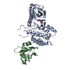
| ||||||||
|---|---|---|---|---|---|---|---|---|---|
| 1 |
| ||||||||
| Unit cell |
|
-Components
| #1: Protein | Mass: 38183.551 Da / Num. of mol.: 1 / Fragment: KINASE DOMAIN, RESIDUES 14-351 Source method: isolated from a genetically manipulated source Source: (gene. exp.) Homo sapiens (human) / Gene: GAK / Plasmid: PNIC28-BSA4 / Production host:  References: UniProt: O14976, non-specific serine/threonine protein kinase |
|---|---|
| #2: Protein | Mass: 15085.511 Da / Num. of mol.: 1 Source method: isolated from a genetically manipulated source Source: (gene. exp.) |
| #3: Chemical | ChemComp-FEF / (  Mass: 365.383 Da / Num. of mol.: 1 / Source method: obtained synthetically / Formula: C20H19N3O4 Mass: 365.383 Da / Num. of mol.: 1 / Source method: obtained synthetically / Formula: C20H19N3O4 |
| #4: Water | ChemComp-HOH /  Mass: 18.015 Da / Num. of mol.: 63 / Source method: isolated from a natural source / Formula: H2O Mass: 18.015 Da / Num. of mol.: 63 / Source method: isolated from a natural source / Formula: H2O |
| Has protein modification | Y |
| Sequence details | SYNTHETIC, NON-BIOLOGICAL |
-Experimental details
-Experiment
| Experiment | Method: X-RAY DIFFRACTION / Number of used crystals: 1 |
|---|
- Sample preparation
| Crystal | Density Matthews: 2.7 Å3/Da / Density % sol: 54 % / Description: NONE |
|---|---|
| Crystal grow | pH: 7.5 / Details: 9% PEG 3350, 0.06 M MAGNESIUM FORMATE, pH 7.5 |
-Data collection
| Diffraction | Mean temperature: 100 K |
|---|---|
| Diffraction source | Source: SYNCHROTRON / Site: Diamond  / Beamline: I04 / Wavelength: 1.0121 / Beamline: I04 / Wavelength: 1.0121 |
| Detector | Type: ADSC QUANTUM 315 / Detector: CCD / Date: Feb 11, 2012 / Details: MIRRORS |
| Radiation | Monochromator: SI (111) DOUBLE CRYSTAL MONOCHROMATOR / Protocol: SINGLE WAVELENGTH / Monochromatic (M) / Laue (L): M / Scattering type: x-ray |
| Radiation wavelength | Wavelength: 1.0121 Å / Relative weight: 1 |
| Reflection | Resolution: 2.8→19.75 Å / Num. obs: 11580 / % possible obs: 99.5 % / Observed criterion σ(I): 2 / Redundancy: 3.6 % / Rmerge(I) obs: 0.14 / Net I/σ(I): 8.3 |
| Reflection shell | Resolution: 2.8→2.95 Å / Redundancy: 3.7 % / Rmerge(I) obs: 0.69 / Mean I/σ(I) obs: 2 / % possible all: 99.8 |
- Processing
| Software |
| ||||||||||||||||||||||||||||||||||||||||||||||||||||||||||||||||||||||||||||||||||||||||||||||||||||||||||||||||||||||||||||||||||||||||||||||||||||||||||||||||||||||||||||||||||||||
|---|---|---|---|---|---|---|---|---|---|---|---|---|---|---|---|---|---|---|---|---|---|---|---|---|---|---|---|---|---|---|---|---|---|---|---|---|---|---|---|---|---|---|---|---|---|---|---|---|---|---|---|---|---|---|---|---|---|---|---|---|---|---|---|---|---|---|---|---|---|---|---|---|---|---|---|---|---|---|---|---|---|---|---|---|---|---|---|---|---|---|---|---|---|---|---|---|---|---|---|---|---|---|---|---|---|---|---|---|---|---|---|---|---|---|---|---|---|---|---|---|---|---|---|---|---|---|---|---|---|---|---|---|---|---|---|---|---|---|---|---|---|---|---|---|---|---|---|---|---|---|---|---|---|---|---|---|---|---|---|---|---|---|---|---|---|---|---|---|---|---|---|---|---|---|---|---|---|---|---|---|---|---|---|
| Refinement | Method to determine structure: MOLECULAR REPLACEMENT Starting model: PDB ENTRIES 3LL6 AND 1OP9 Resolution: 2.8→19.75 Å / Cor.coef. Fo:Fc: 0.935 / Cor.coef. Fo:Fc free: 0.877 / SU B: 32.781 / SU ML: 0.32 / Cross valid method: THROUGHOUT / ESU R Free: 0.394 / Stereochemistry target values: MAXIMUM LIKELIHOOD / Details: HYDROGENS HAVE BEEN ADDED IN THE RIDING POSITIONS.
| ||||||||||||||||||||||||||||||||||||||||||||||||||||||||||||||||||||||||||||||||||||||||||||||||||||||||||||||||||||||||||||||||||||||||||||||||||||||||||||||||||||||||||||||||||||||
| Solvent computation | Ion probe radii: 0.8 Å / Shrinkage radii: 0.8 Å / VDW probe radii: 1.2 Å / Solvent model: MASK | ||||||||||||||||||||||||||||||||||||||||||||||||||||||||||||||||||||||||||||||||||||||||||||||||||||||||||||||||||||||||||||||||||||||||||||||||||||||||||||||||||||||||||||||||||||||
| Displacement parameters | Biso mean: 60.577 Å2
| ||||||||||||||||||||||||||||||||||||||||||||||||||||||||||||||||||||||||||||||||||||||||||||||||||||||||||||||||||||||||||||||||||||||||||||||||||||||||||||||||||||||||||||||||||||||
| Refinement step | Cycle: LAST / Resolution: 2.8→19.75 Å
| ||||||||||||||||||||||||||||||||||||||||||||||||||||||||||||||||||||||||||||||||||||||||||||||||||||||||||||||||||||||||||||||||||||||||||||||||||||||||||||||||||||||||||||||||||||||
| Refine LS restraints |
|