 Movie
Movie Controller

[English] 日本語
 Yorodumi
- PDB-4c1d: Crystal structure of the metallo-beta-lactamase VIM-2 with L-captopril -
+ Open data

- Basic information
| Entry | Database: PDB / ID: 4c1d | ||||||
|---|---|---|---|---|---|---|---|
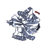
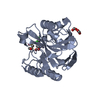
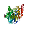
| Title | Crystal structure of the metallo-beta-lactamase VIM-2 with L-captopril | ||||||
 Components Components | BETA-LACTAMASE CLASS B VIM-2 | ||||||
 Keywords Keywords | HYDROLASE / ANTIBIOTIC RESISTANCE | ||||||
| Function / homology |  Function and homology information Function and homology informationantibiotic catabolic process / beta-lactamase activity / beta-lactamase / periplasmic space / response to antibiotic / metal ion binding Similarity search - Function | ||||||
| Biological species |   PSEUDOMONAS AERUGINOSA (bacteria) PSEUDOMONAS AERUGINOSA (bacteria) | ||||||
| Method |  X-RAY DIFFRACTION / SYNCHROTRON / MOLECULAR REPLACEMENT / Resolution: 1.198 Å X-RAY DIFFRACTION / SYNCHROTRON / MOLECULAR REPLACEMENT / Resolution: 1.198 Å | ||||||
 Authors Authors | Zollman, D. / Brem, J. / McDonough, M.A. / van Berkel, S.S. / Schofield, C.J. | ||||||
 Citation Citation | Journal: Antimicrob. Agents Chemother. / Year: 2015 Title: Structural Basis of Metallo-beta-Lactamase Inhibition by Captopril Stereoisomers. Authors: Brem, J. / van Berkel, S.S. / Zollman, D. / Lee, S.Y. / Gileadi, O. / McHugh, P.J. / Walsh, T.R. / McDonough, M.A. / Schofield, C.J. | ||||||
| History |
|
- Structure visualization
| Structure viewer | Molecule: MolmilJmol/JSmol |
|---|
- Downloads & links
-Download
| PDBx/mmCIF format | 4c1d.cif.gz | 211.8 KB | Display | PDBx/mmCIF format |
|---|---|---|---|---|
| PDB format | pdb4c1d.ent.gz | 169.2 KB | Display | PDB format |
| PDBx/mmJSON format | 4c1d.json.gz | Tree view | PDBx/mmJSON format | |
| Others |  Other downloads Other downloads |
-Validation report
| Arichive directory | https://data.pdbj.org/pub/pdb/validation_reports/c1/4c1dftp://data.pdbj.org/pub/pdb/validation_reports/c1/4c1d | HTTPS FTP |
|---|
-Related structure data
| Related structure data |  4bz3C  4c09C  4c1cC 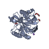 4c1eC  4c1fC  4c1gC  4c1hC  1ko3S C: citing same article ( S: Starting model for refinement |
|---|---|
| Similar structure data |


-Links
 PDBj
PDBj

- Assembly
| Deposited unit | 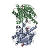
| ||||||||
|---|---|---|---|---|---|---|---|---|---|
| 1 | 
| ||||||||
| 2 | 
| ||||||||
| Unit cell |
|
-Components
-Protein , 1 types, 2 molecules AB
| #1: Protein | Mass: 25693.488 Da / Num. of mol.: 2 / Fragment: RESIDUES 2-240 Source method: isolated from a genetically manipulated source Source: (gene. exp.) PSEUDOMONAS AERUGINOSA (bacteria) / Description: PLASMID DERIVED NON-GENOMIC. / Plasmid: OPINF VECTOR BASED ON PTRIEX VECTOR / Production host: |
|---|
-Non-polymers , 5 types, 446 molecules 


| #2: Chemical |  Mass: 22.990 Da / Num. of mol.: 2 / Source method: obtained synthetically / Formula: Na Mass: 22.990 Da / Num. of mol.: 2 / Source method: obtained synthetically / Formula: Na#3: Chemical |  Mass: 217.285 Da / Num. of mol.: 2 / Source method: obtained synthetically / Formula: C9H15NO3S / Comment: inhibitor, medication*YM Mass: 217.285 Da / Num. of mol.: 2 / Source method: obtained synthetically / Formula: C9H15NO3S / Comment: inhibitor, medication*YM#4: Chemical | ChemComp-FMT /  Mass: 46.025 Da / Num. of mol.: 4 / Source method: obtained synthetically / Formula: CH2O2 Mass: 46.025 Da / Num. of mol.: 4 / Source method: obtained synthetically / Formula: CH2O2#5: Chemical | ChemComp-ZN /  Mass: 65.409 Da / Num. of mol.: 6 / Source method: obtained synthetically / Formula: Zn Mass: 65.409 Da / Num. of mol.: 6 / Source method: obtained synthetically / Formula: Zn#6: Water | ChemComp-HOH / | Mass: 18.015 Da / Num. of mol.: 432 / Source method: isolated from a natural source / Formula: H2O |
|---|
-Experimental details
-Experiment
| Experiment | Method: X-RAY DIFFRACTION / Number of used crystals: 1 |
|---|
- Sample preparation
| Crystal | Density Matthews: 2.32 Å3/Da / Density % sol: 47 % / Description: NONE |
|---|---|
| Crystal grow | Details: 0.2 M MAGNESIUM FORMATE, 20 % W/V PEG3350, 1 MM TCEP. |
-Data collection
| Diffraction | Mean temperature: 100 K |
|---|---|
| Diffraction source | Source: SYNCHROTRON / Site: Diamond  / Beamline: I03 / Wavelength: 0.9763 / Beamline: I03 / Wavelength: 0.9763 |
| Detector | Type: DECTRIS PILATUS / Detector: PIXEL / Date: Oct 22, 2012 |
| Radiation | Protocol: SINGLE WAVELENGTH / Monochromatic (M) / Laue (L): M / Scattering type: x-ray |
| Radiation wavelength | Wavelength: 0.9763 Å / Relative weight: 1 |
| Reflection | Resolution: 1.2→50 Å / Num. obs: 126706 / % possible obs: 99.9 % / Observed criterion σ(I): 0 / Redundancy: 4.6 % / Biso Wilson estimate: 13.24 Å2 / Rmerge(I) obs: 0.17 / Net I/σ(I): 10.6 |
| Reflection shell | Resolution: 1.2→1.24 Å / Redundancy: 2.5 % / Rmerge(I) obs: 0.61 / Mean I/σ(I) obs: 1.9 / % possible all: 91 |
- Processing
| Software |
| |||||||||||||||||||||||||||||||||||||||||||||||||||||||||||||||||||||||||||||||||||||||||||||||||||||||||
|---|---|---|---|---|---|---|---|---|---|---|---|---|---|---|---|---|---|---|---|---|---|---|---|---|---|---|---|---|---|---|---|---|---|---|---|---|---|---|---|---|---|---|---|---|---|---|---|---|---|---|---|---|---|---|---|---|---|---|---|---|---|---|---|---|---|---|---|---|---|---|---|---|---|---|---|---|---|---|---|---|---|---|---|---|---|---|---|---|---|---|---|---|---|---|---|---|---|---|---|---|---|---|---|---|---|---|
| Refinement | Method to determine structure: MOLECULAR REPLACEMENT Starting model: PDB ENTRY 1KO3 Resolution: 1.198→39.701 Å / SU ML: 0.13 / σ(F): 1.34 / Phase error: 18.89 / Stereochemistry target values: ML Details: CONFORMATION OF RAMACHANDRAN OUTLIERS 84 ASP, 87 TRP AND 178 ALA VALIDATED BY CLEAR ELECTRON DENSITY. POSSIBLE ALTERNATIVE CONFORMATION OF LIGAND, HOWEVER VERY LOW OCCUPANCY SO LEFT UNMODELLED.
| |||||||||||||||||||||||||||||||||||||||||||||||||||||||||||||||||||||||||||||||||||||||||||||||||||||||||
| Solvent computation | Shrinkage radii: 0.9 Å / VDW probe radii: 1.11 Å / Solvent model: FLAT BULK SOLVENT MODEL | |||||||||||||||||||||||||||||||||||||||||||||||||||||||||||||||||||||||||||||||||||||||||||||||||||||||||
| Displacement parameters | Biso mean: 18.82 Å2 | |||||||||||||||||||||||||||||||||||||||||||||||||||||||||||||||||||||||||||||||||||||||||||||||||||||||||
| Refinement step | Cycle: LAST / Resolution: 1.198→39.701 Å
| |||||||||||||||||||||||||||||||||||||||||||||||||||||||||||||||||||||||||||||||||||||||||||||||||||||||||
| Refine LS restraints |
| |||||||||||||||||||||||||||||||||||||||||||||||||||||||||||||||||||||||||||||||||||||||||||||||||||||||||
| LS refinement shell |
|
