 Movie
Movie Controller

[English] 日本語
 Yorodumi
- PDB-1ko3: VIM-2, a Zn-beta-lactamase from Pseudomonas aeruginosa with Cys22... -
+ Open data

- Basic information
| Entry | Database: PDB / ID: 1ko3 | ||||||
|---|---|---|---|---|---|---|---|
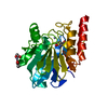
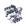
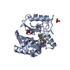
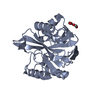
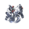
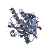
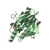
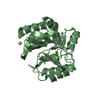
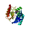
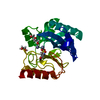
| Title | VIM-2, a Zn-beta-lactamase from Pseudomonas aeruginosa with Cys221 reduced | ||||||
 Components Components | VIM-2 metallo-beta-lactamase | ||||||
 Keywords Keywords | HYDROLASE / alpha-beta/beta-alpha fold | ||||||
| Function / homology |  Function and homology information Function and homology informationantibiotic catabolic process / beta-lactamase / beta-lactamase activity / periplasmic space / response to antibiotic / metal ion binding Similarity search - Function | ||||||
| Biological species |   Pseudomonas aeruginosa (bacteria) Pseudomonas aeruginosa (bacteria) | ||||||
| Method |  X-RAY DIFFRACTION / MOLECULAR REPLACEMENT / Resolution: 1.91 Å X-RAY DIFFRACTION / MOLECULAR REPLACEMENT / Resolution: 1.91 Å | ||||||
 Authors Authors | Garcia-Saez, I. / Docquier, J.-D. / Rossolini, G.M. / Dideberg, O. | ||||||
 Citation Citation | Journal: J.Mol.Biol. / Year: 2008 Title: The three-dimensional structure of VIM-2, a Zn-beta-lactamase from Pseudomonas aeruginosa in its reduced and oxidised form Authors: Garcia-Saez, I. / Docquier, J.-D. / Rossolini, G.M. / Dideberg, O. | ||||||
| History |
|
- Structure visualization
| Structure viewer | Molecule: MolmilJmol/JSmol |
|---|
- Downloads & links
-Download
| PDBx/mmCIF format | 1ko3.cif.gz | 60.8 KB | Display | PDBx/mmCIF format |
|---|---|---|---|---|
| PDB format | pdb1ko3.ent.gz | 42.9 KB | Display | PDB format |
| PDBx/mmJSON format | 1ko3.json.gz | Tree view | PDBx/mmJSON format | |
| Others |  Other downloads Other downloads |
-Validation report
| Arichive directory | https://data.pdbj.org/pub/pdb/validation_reports/ko/1ko3ftp://data.pdbj.org/pub/pdb/validation_reports/ko/1ko3 | HTTPS FTP |
|---|
-Related structure data
| Related structure data |  1ko2SC S: Starting model for refinement C: citing same article ( |
|---|---|
| Similar structure data |


-Links
 PDBj
PDBj


- Assembly
| Deposited unit | 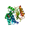
| ||||||||
|---|---|---|---|---|---|---|---|---|---|
| 1 |
| ||||||||
| Unit cell |
|
-Components
-Protein , 1 types, 1 molecules A
| #1: Protein | Mass: 24522.246 Da / Num. of mol.: 1 / Fragment: residues 30-295 Source method: isolated from a genetically manipulated source Source: (gene. exp.) Pseudomonas aeruginosa (bacteria) / Gene: blaVIM-2 / Plasmid: PET9-VIM-2 / Species (production host): Escherichia coli / Production host: |
|---|
-Non-polymers , 5 types, 143 molecules 


| #2: Chemical |  Mass: 65.409 Da / Num. of mol.: 3 / Source method: obtained synthetically / Formula: Zn Mass: 65.409 Da / Num. of mol.: 3 / Source method: obtained synthetically / Formula: Zn#3: Chemical |  Mass: 59.044 Da / Num. of mol.: 2 / Source method: obtained synthetically / Formula: C2H3O2 Mass: 59.044 Da / Num. of mol.: 2 / Source method: obtained synthetically / Formula: C2H3O2#4: Chemical | ChemComp-CL / |  Mass: 35.453 Da / Num. of mol.: 1 / Source method: obtained synthetically / Formula: Cl Mass: 35.453 Da / Num. of mol.: 1 / Source method: obtained synthetically / Formula: Cl#5: Chemical | ChemComp-OH / |  Mass: 17.007 Da / Num. of mol.: 1 / Source method: obtained synthetically / Formula: HO Mass: 17.007 Da / Num. of mol.: 1 / Source method: obtained synthetically / Formula: HO#6: Water | ChemComp-HOH / | Mass: 18.015 Da / Num. of mol.: 136 / Source method: isolated from a natural source / Formula: H2O |
|---|
-Details
| Sequence details | THIS COORDINATES IS USED NON-SEQUENTIAL RESIDUE NUMBERING. MANY NUMBERS WERE SIMPLY SKIPPED IN THE ...THIS COORDINATE |
|---|
-Experimental details
-Experiment
| Experiment | Method: X-RAY DIFFRACTION / Number of used crystals: 1 |
|---|
- Sample preparation
| Crystal | Density Matthews: 2.15 Å3/Da / Density % sol: 42.72 % | |||||||||||||||||||||||||||||||||||||||||||||||||||||||||||||||
|---|---|---|---|---|---|---|---|---|---|---|---|---|---|---|---|---|---|---|---|---|---|---|---|---|---|---|---|---|---|---|---|---|---|---|---|---|---|---|---|---|---|---|---|---|---|---|---|---|---|---|---|---|---|---|---|---|---|---|---|---|---|---|---|---|
| Crystal grow | Temperature: 293.15 K / Method: vapor diffusion, hanging drop / pH: 6.5 Details: 30% PEG8K, 0.1M NaCac/Cac.acid, 0.2M Na acetate, 0.5mM TCEP, pH 6.5, VAPOR DIFFUSION, HANGING DROP, temperature 293.15K | |||||||||||||||||||||||||||||||||||||||||||||||||||||||||||||||
| Crystal grow | *PLUS Temperature: 20 ℃ / pH: 7.5 / Method: vapor diffusion, hanging drop | |||||||||||||||||||||||||||||||||||||||||||||||||||||||||||||||
| Components of the solutions | *PLUS
|
-Data collection
| Diffraction | Mean temperature: 100 K |
|---|---|
| Diffraction source | Source: ROTATING ANODE / Type: ENRAF-NONIUS FR591 / Wavelength: 1.5418 Å |
| Detector | Type: MARRESEARCH / Detector: IMAGE PLATE / Date: Oct 24, 2001 / Details: mirrors |
| Radiation | Protocol: SINGLE WAVELENGTH / Monochromatic (M) / Laue (L): M / Scattering type: x-ray |
| Radiation wavelength | Wavelength: 1.5418 Å / Relative weight: 1 |
| Reflection | Resolution: 1.91→51.63 Å / Num. all: 16087 / Num. obs: 15030 / % possible obs: 90.6 % / Redundancy: 3.6 % / Rmerge(I) obs: 0.052 / Rsym value: 0.045 / Net I/σ(I): 12.6 |
| Reflection shell | Resolution: 1.91→2.02 Å / Redundancy: 1.7 % / Rmerge(I) obs: 0.171 / Mean I/σ(I) obs: 5.4 / Num. unique all: 544 / Rsym value: 0.135 / % possible all: 50 |
| Reflection | *PLUS Highest resolution: 1.9 Å / % possible obs: 91 % / Rmerge(I) obs: 0.05 |
| Reflection shell | *PLUS % possible obs: 50 % / Rmerge(I) obs: 0.14 |
- Processing
| Software |
| ||||||||||||||||||||||||||||
|---|---|---|---|---|---|---|---|---|---|---|---|---|---|---|---|---|---|---|---|---|---|---|---|---|---|---|---|---|---|
| Refinement | Method to determine structure: MOLECULAR REPLACEMENT Starting model: VIM-2 oxidized, PDB ID code 1KO2 Resolution: 1.91→25 Å / Isotropic thermal model: restrained / Cross valid method: THROUGHOUT / σ(F): 0
| ||||||||||||||||||||||||||||
| Refine analyze |
| ||||||||||||||||||||||||||||
| Refinement step | Cycle: LAST / Resolution: 1.91→25 Å
| ||||||||||||||||||||||||||||
| Refine LS restraints |
| ||||||||||||||||||||||||||||
| LS refinement shell |
| ||||||||||||||||||||||||||||
| Refinement | *PLUS Highest resolution: 1.9 Å / % reflection Rfree: 10 % / Rfactor Rfree: 0.25 / Rfactor Rwork: 0.209 | ||||||||||||||||||||||||||||
| Solvent computation | *PLUS | ||||||||||||||||||||||||||||
| Displacement parameters | *PLUS | ||||||||||||||||||||||||||||
| Refine LS restraints | *PLUS
| ||||||||||||||||||||||||||||
| LS refinement shell | *PLUS Lowest resolution: 2.02 Å |
