 Movie
Movie Controller


+ Open data

- Basic information
| Entry | Database: PDB / ID: 4bnq | ||||||
|---|---|---|---|---|---|---|---|
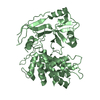
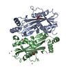
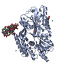
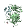
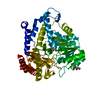
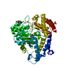
| Title | The structure of the Staphylococcus aureus Ham1 protein | ||||||
 Components Components | NON-CANONICAL PURINE NTP PYROPHOSPHATASE | ||||||
 Keywords Keywords | HYDROLASE / HAM / INOSINE TRIPHOSPHATE PYROPHOSPHATASE | ||||||
| Function / homology |  Function and homology information Function and homology informationpurine nucleoside triphosphate catabolic process / XTP/dITP diphosphatase / ITP diphosphatase activity / XTP diphosphatase activity / dITP diphosphatase activity / nucleotide metabolic process / ribonucleoside triphosphate phosphatase activity / nucleotide binding / metal ion binding / cytosol Similarity search - Function | ||||||
| Biological species |   STAPHYLOCOCCUS AUREUS SUBSP. AUREUS (bacteria) STAPHYLOCOCCUS AUREUS SUBSP. AUREUS (bacteria) | ||||||
| Method |  X-RAY DIFFRACTION / SYNCHROTRON / MAD / Resolution: 2.279 Å X-RAY DIFFRACTION / SYNCHROTRON / MAD / Resolution: 2.279 Å | ||||||
 Authors Authors | Abergel, C. / Claverie, J.M. | ||||||
 Citation Citation | Journal: Acta Crystallogr.,Sect.D / Year: 2013 Title: Molecular Replacement: Tricks and Treats. Authors: Abergel, C. | ||||||
| History |
|
- Structure visualization
| Structure viewer | Molecule: MolmilJmol/JSmol |
|---|
- Downloads & links
-Download
| PDBx/mmCIF format | 4bnq.cif.gz | 89 KB | Display | PDBx/mmCIF format |
|---|---|---|---|---|
| PDB format | pdb4bnq.ent.gz | 69.8 KB | Display | PDB format |
| PDBx/mmJSON format | 4bnq.json.gz | Tree view | PDBx/mmJSON format | |
| Others |  Other downloads Other downloads |
-Validation report
| Arichive directory | https://data.pdbj.org/pub/pdb/validation_reports/bn/4bnqftp://data.pdbj.org/pub/pdb/validation_reports/bn/4bnq | HTTPS FTP |
|---|
-Related structure data
| Similar structure data |
|---|


-Links
 PDBj
PDBj- Assembly
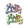
| Deposited unit | 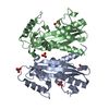
| ||||||||
|---|---|---|---|---|---|---|---|---|---|
| 1 |
| ||||||||
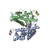
| Unit cell |
|
-Components
| #1: Protein | Mass: 21441.094 Da / Num. of mol.: 2 Source method: isolated from a genetically manipulated source Source: (gene. exp.) STAPHYLOCOCCUS AUREUS SUBSP. AUREUS (bacteria)Strain: N315 / Plasmid: IN HOUSE / Production host: References: UniProt: P99094, nucleoside-triphosphate diphosphatase #2: Chemical | ChemComp-PO4 /   Mass: 94.971 Da / Num. of mol.: 5 / Source method: obtained synthetically / Formula: PO4 Mass: 94.971 Da / Num. of mol.: 5 / Source method: obtained synthetically / Formula: PO4#3: Chemical | ChemComp-GOL / |   Mass: 92.094 Da / Num. of mol.: 1 / Source method: obtained synthetically / Formula: C3H8O3 Mass: 92.094 Da / Num. of mol.: 1 / Source method: obtained synthetically / Formula: C3H8O3#4: Water | ChemComp-HOH / |  Mass: 18.015 Da / Num. of mol.: 193 / Source method: isolated from a natural source / Formula: H2O Mass: 18.015 Da / Num. of mol.: 193 / Source method: isolated from a natural source / Formula: H2OSequence details | LACK OF INITIAL MET | |
|---|
-Experimental details
-Experiment
| Experiment | Method: X-RAY DIFFRACTION / Number of used crystals: 1 |
|---|
- Sample preparation
| Crystal | Density Matthews: 3.31 Å3/Da / Density % sol: 63 % / Description: NONE |
|---|---|
| Crystal grow | Details: 20% AMSO4, PH 6 |
-Data collection
| Diffraction | Mean temperature: 105 K |
|---|---|
| Diffraction source | Source: SYNCHROTRON / Site: ESRF  / Beamline: BM30A / Wavelength: 0.97974 / Beamline: BM30A / Wavelength: 0.97974 |
| Detector | Type: ADSC QUANTUM 315 / Detector: CCD |
| Radiation | Protocol: SINGLE WAVELENGTH / Monochromatic (M) / Laue (L): M / Scattering type: x-ray |
| Radiation wavelength | Wavelength: 0.97974 Å / Relative weight: 1 |
| Reflection | Resolution: 2.28→43.4 Å / Num. obs: 26528 / % possible obs: 85.7 % / Observed criterion σ(I): 1.34 / Redundancy: 4.1 % / Biso Wilson estimate: 34.8 Å2 / Rmerge(I) obs: 0.09 / Net I/σ(I): 5.8 |
| Reflection shell | Resolution: 2.28→2.4 Å / Redundancy: 2.4 % / Rmerge(I) obs: 0.19 / Mean I/σ(I) obs: 3.4 / % possible all: 85.7 |
- Processing
| Software |
| |||||||||||||||||||||||||||||||||||||||||||||||||||||||||||||||
|---|---|---|---|---|---|---|---|---|---|---|---|---|---|---|---|---|---|---|---|---|---|---|---|---|---|---|---|---|---|---|---|---|---|---|---|---|---|---|---|---|---|---|---|---|---|---|---|---|---|---|---|---|---|---|---|---|---|---|---|---|---|---|---|---|
| Refinement | Method to determine structure: MAD Starting model: NONE Resolution: 2.279→43.398 Å / SU ML: 0.27 / σ(F): 1.34 / Phase error: 25.46 / Stereochemistry target values: ML / Details: THE TWO MONOMERS ADOPT DIFFERENT CONFORMATIONS
| |||||||||||||||||||||||||||||||||||||||||||||||||||||||||||||||
| Solvent computation | Shrinkage radii: 0.9 Å / VDW probe radii: 1.11 Å / Solvent model: FLAT BULK SOLVENT MODEL | |||||||||||||||||||||||||||||||||||||||||||||||||||||||||||||||
| Refinement step | Cycle: LAST / Resolution: 2.279→43.398 Å
| |||||||||||||||||||||||||||||||||||||||||||||||||||||||||||||||
| Refine LS restraints |
| |||||||||||||||||||||||||||||||||||||||||||||||||||||||||||||||
| LS refinement shell |
|
