 Movie
Movie Controller

[English] 日本語
 Yorodumi
- PDB-4zlt: Crystal structure of viral chemokine binding protein R17 in compl... -
+ Open data

- Basic information
| Entry | Database: PDB / ID: 4zlt | |||||||||||||||
|---|---|---|---|---|---|---|---|---|---|---|---|---|---|---|---|---|
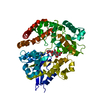
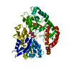
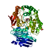
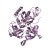
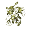
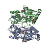
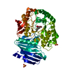
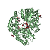
| Title | Crystal structure of viral chemokine binding protein R17 in complex with CCL3 | |||||||||||||||
 Components Components |
| |||||||||||||||
 Keywords Keywords | Chemokine binding protein/Chemokine / RHVP chemokine binding protein in complex with chemokine CCL3 / Chemokine binding protein-Chemokine complex | |||||||||||||||
| Function / homology |  Function and homology information Function and homology informationChemokine receptors bind chemokines / lymphocyte chemotaxis / granulocyte chemotaxis / positive regulation of natural killer cell chemotaxis / astrocyte cell migration / eosinophil degranulation / regulation of sensory perception of pain / signaling / cell activation / T cell chemotaxis ...Chemokine receptors bind chemokines / lymphocyte chemotaxis / granulocyte chemotaxis / positive regulation of natural killer cell chemotaxis / astrocyte cell migration / eosinophil degranulation / regulation of sensory perception of pain / signaling / cell activation / T cell chemotaxis / positive regulation of calcium ion transport / eosinophil chemotaxis / positive regulation of osteoclast differentiation / response to cholesterol / chemokine activity / release of sequestered calcium ion into cytosol by sarcoplasmic reticulum / negative regulation of osteoclast differentiation / phospholipase activator activity / chemoattractant activity / exocytosis / macrophage chemotaxis / monocyte chemotaxis / host-mediated suppression of viral transcription / neutrophil chemotaxis / positive regulation of calcium-mediated signaling / cytoskeleton organization / positive regulation of interleukin-1 beta production / calcium-mediated signaling / response to toxic substance / intracellular calcium ion homeostasis / chemotaxis / osteoblast differentiation / kinase activity / calcium ion transport / positive regulation of inflammatory response / positive regulation of tumor necrosis factor production / positive regulation of neuron apoptotic process / cell-cell signaling / regulation of cell shape / positive regulation of cytosolic calcium ion concentration / protein phosphorylation / positive regulation of ERK1 and ERK2 cascade / protein kinase activity / positive regulation of cell migration / response to xenobiotic stimulus / inflammatory response / negative regulation of gene expression / positive regulation of gene expression / positive regulation of transcription by RNA polymerase II / : / cytosol / cytoplasm Similarity search - Function | |||||||||||||||
| Biological species |  Cricetid herpesvirus 2 Cricetid herpesvirus 2 | |||||||||||||||
| Method |  X-RAY DIFFRACTION / SYNCHROTRON / MOLECULAR REPLACEMENT / Resolution: 3 Å X-RAY DIFFRACTION / SYNCHROTRON / MOLECULAR REPLACEMENT / Resolution: 3 Å | |||||||||||||||
 Authors Authors | Lubman, O.Y. / Fremont, D.H. | |||||||||||||||
| Funding support |  United States, 4items United States, 4items
| |||||||||||||||
 Citation Citation | Journal: Structure / Year: 2016 Title: Parallel Evolution of Chemokine Binding by Structurally Related Herpesvirus Decoy Receptors. Authors: Lubman, O.Y. / Fremont, D.H. | |||||||||||||||
| History |
|
- Structure visualization
| Structure viewer | Molecule: MolmilJmol/JSmol |
|---|
- Downloads & links
-Download
| PDBx/mmCIF format | 4zlt.cif.gz | 187.1 KB | Display | PDBx/mmCIF format |
|---|---|---|---|---|
| PDB format | pdb4zlt.ent.gz | 148.9 KB | Display | PDB format |
| PDBx/mmJSON format | 4zlt.json.gz | Tree view | PDBx/mmJSON format | |
| Others |  Other downloads Other downloads |
-Validation report
| Arichive directory | https://data.pdbj.org/pub/pdb/validation_reports/zl/4zltftp://data.pdbj.org/pub/pdb/validation_reports/zl/4zlt | HTTPS FTP |
|---|
-Related structure data
| Related structure data |  4zkqSC  2x6gS S: Starting model for refinement C: citing same article ( |
|---|---|
| Similar structure data |


-Links
 PDBj
PDBj
- Assembly
| Deposited unit | 
| ||||||||
|---|---|---|---|---|---|---|---|---|---|
| 1 | 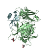
| ||||||||
| 2 | 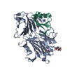
| ||||||||
| Unit cell |
| ||||||||
| Details | dimer according to multi-angle static light scattering |
-Components
| #1: Protein | Mass: 47388.625 Da / Num. of mol.: 2 / Mutation: K333D, R335E, R336E, K337D Source method: isolated from a genetically manipulated source Source: (gene. exp.) Cricetid herpesvirus 2 / Gene: RHVP-L.R17, RHVP.R17 / Cell (production host): endothelial / Cell line (production host): 293F / Production host: Mammalian expression vector pBGSA (others) / References: UniProt: E9M5R0#2: Protein | Mass: 7982.065 Da / Num. of mol.: 2 / Mutation: D27A Source method: isolated from a genetically manipulated source Source: (gene. exp.)  #3: Sugar | ChemComp-NAG /   Type: D-saccharide, beta linking / Mass: 221.208 Da / Num. of mol.: 4 Type: D-saccharide, beta linking / Mass: 221.208 Da / Num. of mol.: 4Source method: isolated from a genetically manipulated source Formula: C8H15NO6 Has protein modification | Y | |
|---|
-Experimental details
-Experiment
| Experiment | Method: X-RAY DIFFRACTION |
|---|
- Sample preparation
| Crystal | Density Matthews: 2.57 Å3/Da / Density % sol: 52.1 % |
|---|---|
| Crystal grow | Temperature: 297 K / Method: vapor diffusion, hanging drop / Details: 15-20% PEG 3350 0.2-0.4M MgFormate |
-Data collection
| Diffraction | Mean temperature: 100 K | |||||||||
|---|---|---|---|---|---|---|---|---|---|---|
| Diffraction source | Source: SYNCHROTRON / Site: ALS / Beamline: 4.2.2 / Wavelength: 1 Å | |||||||||
| Detector | Type: NOIR-1 / Detector: CCD / Date: Jun 13, 2013 | |||||||||
| Radiation | Protocol: SINGLE WAVELENGTH / Monochromatic (M) / Laue (L): M / Scattering type: x-ray | |||||||||
| Radiation wavelength | Wavelength: 1 Å / Relative weight: 1 | |||||||||
| Reflection | Resolution: 2.761→50 Å / Num. obs: 26825 / % possible obs: 100 % / Redundancy: 4.7 % / Rsym value: 0.11 / Net I/σ(I): 7.4 | |||||||||
| Reflection shell |
|
- Processing
| Software |
| |||||||||||||||||||||||||||||||||||||||||||||||||||||||||||||||||||||||||||||||||||||||||||
|---|---|---|---|---|---|---|---|---|---|---|---|---|---|---|---|---|---|---|---|---|---|---|---|---|---|---|---|---|---|---|---|---|---|---|---|---|---|---|---|---|---|---|---|---|---|---|---|---|---|---|---|---|---|---|---|---|---|---|---|---|---|---|---|---|---|---|---|---|---|---|---|---|---|---|---|---|---|---|---|---|---|---|---|---|---|---|---|---|---|---|---|---|
| Refinement | Method to determine structure: MOLECULAR REPLACEMENT Starting model: 4ZKQ and 2X6G Resolution: 3→49.247 Å / SU ML: 0.4 / Cross valid method: FREE R-VALUE / σ(F): 0 / Phase error: 27.94 / Stereochemistry target values: ML
| |||||||||||||||||||||||||||||||||||||||||||||||||||||||||||||||||||||||||||||||||||||||||||
| Solvent computation | Shrinkage radii: 0.9 Å / VDW probe radii: 1.11 Å / Solvent model: FLAT BULK SOLVENT MODEL | |||||||||||||||||||||||||||||||||||||||||||||||||||||||||||||||||||||||||||||||||||||||||||
| Refinement step | Cycle: LAST / Resolution: 3→49.247 Å
| |||||||||||||||||||||||||||||||||||||||||||||||||||||||||||||||||||||||||||||||||||||||||||
| Refine LS restraints |
| |||||||||||||||||||||||||||||||||||||||||||||||||||||||||||||||||||||||||||||||||||||||||||
| LS refinement shell |
|
