[English] 日本語
Yorodumi
- PDB-3vfj: The structure of monodechloro-teicoplanin in complex with its lig... -

+
Open data


ID or keywords:

Loading...

-
Basic information

Entry
Database: PDB / ID: 3vfj
TitleThe structure of monodechloro-teicoplanin in complex with its ligand, using MBP as a ligand carrier
Components
  • Maltose-binding periplasmic protein, C-terminal fused by Cys-Lys-D-Ala-D-Ala
  • MonodeChloro- Teicoplanin A2-2
KeywordsSUGAR BINDING PROTEIN/ANTIBIOTIC / teicoplanin / acetylation of cyteine with iodoacetate modification / SUGAR BINDING PROTEIN-ANTIBIOTIC complex
Function / homology
Function and homology information


detection of maltose stimulus / maltose transport complex / carbohydrate transport / carbohydrate transmembrane transporter activity / maltose binding / maltose transport / maltodextrin transmembrane transport / ATP-binding cassette (ABC) transporter complex, substrate-binding subunit-containing / ATP-binding cassette (ABC) transporter complex / cell chemotaxis ...detection of maltose stimulus / maltose transport complex / carbohydrate transport / carbohydrate transmembrane transporter activity / maltose binding / maltose transport / maltodextrin transmembrane transport / ATP-binding cassette (ABC) transporter complex, substrate-binding subunit-containing / ATP-binding cassette (ABC) transporter complex / cell chemotaxis / outer membrane-bounded periplasmic space / periplasmic space / DNA damage response / membrane
Similarity search - Function
Maltose/Cyclodextrin ABC transporter, substrate-binding protein / Solute-binding family 1, conserved site / Bacterial extracellular solute-binding proteins, family 1 signature. / Bacterial extracellular solute-binding protein / Bacterial extracellular solute-binding protein / Periplasmic binding protein-like II / D-Maltodextrin-Binding Protein; domain 2 / 3-Layer(aba) Sandwich / Alpha Beta
Similarity search - Domain/homology
MonodeChloro- Teicoplanin A2-2 / ACETATE ION / CACODYLATE ION / 2-amino-2-deoxy-beta-D-glucopyranose / alpha-D-mannopyranose / 8-METHYLNONANOIC ACID / Maltose/maltodextrin-binding periplasmic protein
Similarity search - Component
Biological speciesEscherichia coli (E. coli)
Actinoplanes teichomyceticus (bacteria)
MethodX-RAY DIFFRACTION / SYNCHROTRON / MOLECULAR REPLACEMENT / molecular replacement / Resolution: 2.05 Å
AuthorsEconomou, N.J. / Weeks, S.D. / Grasty, K.C. / Loll, P.J.
CitationJournal: Acta Crystallogr.,Sect.D / Year: 2013
Title: Structure of the complex between teicoplanin and a bacterial cell-wall peptide: use of a carrier-protein approach.
Authors: Economou, N.J. / Zentner, I.J. / Lazo, E. / Jakoncic, J. / Stojanoff, V. / Weeks, S.D. / Grasty, K.C. / Cocklin, S. / Loll, P.J.
History
DepositionJan 9, 2012Deposition site: RCSB / Processing site: RCSB
Revision 1.0Jan 9, 2013Provider: repository / Type: Initial release
Revision 1.1Mar 6, 2013Group: Structure summary
Revision 1.2Mar 13, 2013Group: Structure summary
Revision 1.3Mar 27, 2013Group: Derived calculations
Revision 1.4Apr 3, 2013Group: Derived calculations
Revision 1.5Jun 19, 2013Group: Database references
Revision 1.6Dec 10, 2014Group: Structure summary
Revision 2.0Jul 29, 2020Group: Data collection / Database references ...Data collection / Database references / Derived calculations / Polymer sequence / Structure summary
Category: chem_comp / entity ...chem_comp / entity / entity_poly / pdbx_chem_comp_identifier / pdbx_entity_nonpoly / pdbx_struct_conn_angle / struct_conn / struct_ref_seq_dif / struct_site / struct_site_gen
Item: _chem_comp.name / _chem_comp.type ..._chem_comp.name / _chem_comp.type / _entity.pdbx_description / _entity_poly.pdbx_seq_one_letter_code_can / _pdbx_entity_nonpoly.name / _pdbx_struct_conn_angle.ptnr1_auth_comp_id / _pdbx_struct_conn_angle.ptnr1_auth_seq_id / _pdbx_struct_conn_angle.ptnr1_label_asym_id / _pdbx_struct_conn_angle.ptnr1_label_atom_id / _pdbx_struct_conn_angle.ptnr1_label_comp_id / _pdbx_struct_conn_angle.ptnr1_label_seq_id / _pdbx_struct_conn_angle.ptnr2_auth_seq_id / _pdbx_struct_conn_angle.ptnr2_label_asym_id / _pdbx_struct_conn_angle.ptnr3_auth_comp_id / _pdbx_struct_conn_angle.ptnr3_auth_seq_id / _pdbx_struct_conn_angle.ptnr3_label_asym_id / _pdbx_struct_conn_angle.ptnr3_label_atom_id / _pdbx_struct_conn_angle.ptnr3_label_comp_id / _pdbx_struct_conn_angle.ptnr3_label_seq_id / _pdbx_struct_conn_angle.value / _struct_conn.conn_type_id / _struct_conn.id / _struct_conn.pdbx_dist_value / _struct_conn.pdbx_leaving_atom_flag / _struct_conn.ptnr1_auth_asym_id / _struct_conn.ptnr1_auth_comp_id / _struct_conn.ptnr1_auth_seq_id / _struct_conn.ptnr1_label_asym_id / _struct_conn.ptnr1_label_atom_id / _struct_conn.ptnr1_label_comp_id / _struct_conn.ptnr1_label_seq_id / _struct_conn.ptnr2_auth_asym_id / _struct_conn.ptnr2_auth_comp_id / _struct_conn.ptnr2_auth_seq_id / _struct_conn.ptnr2_label_asym_id / _struct_conn.ptnr2_label_atom_id / _struct_conn.ptnr2_label_comp_id / _struct_conn.ptnr2_label_seq_id / _struct_ref_seq_dif.details
Description: Carbohydrate remediation / Provider: repository / Type: Remediation

-
Structure visualization

Structure viewerMolecule:
MolmilJmol/JSmol

Downloads & links

-
Assembly

Deposited unit
A: Maltose-binding periplasmic protein, C-terminal fused by Cys-Lys-D-Ala-D-Ala
G: MonodeChloro- Teicoplanin A2-2
hetero molecules


Theoretical massNumber of molelcules
Total (without water)44,18318
Polymers42,5152
Non-polymers1,66816
Water2,792155
1


  • Idetical with deposited unit
  • defined by author&software
TypeNameSymmetry operationNumber
identity operation1_555x,y,z1
Buried area2930 Å2
ΔGint-154 kcal/mol
Surface area16900 Å2
MethodPISA
Unit cell
Length a, b, c (Å)40.320, 123.610, 156.740
Angle α, β, γ (deg.)90.00, 90.00, 90.00
Int Tables number20
Space group name H-MC2221
Components on special symmetry positions
IDModelComponents
11A-403-

ZN

-
Components

-
Protein / Protein/peptide , 2 types, 2 molecules AG

#1: Protein Maltose-binding periplasmic protein, C-terminal fused by Cys-Lys-D-Ala-D-Ala / MBP / MMBP / Maltodextrin-binding protein


Mass: 41342.777 Da / Num. of mol.: 1
Source method: isolated from a genetically manipulated source
Details: four C-terminal amino acids added non-recombinantly with native chemical ligation
Source: (gene. exp.) Escherichia coli (E. coli) / Strain: K12 / Gene: malE, b4034, JW3994 / Production host: Escherichia coli (E. coli) / References: UniProt: P0AEX9
#2: Protein/peptide MonodeChloro- Teicoplanin A2-2


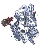
Type: Glycopeptide / Class: Antibiotic / Mass: 1172.539 Da / Num. of mol.: 1 / Source method: isolated from a natural source / Source: (natural) Actinoplanes teichomyceticus (bacteria) / References: MonodeChloro- Teicoplanin A2-2

-
Sugars , 3 types, 3 molecules

#6: Sugar ChemComp-GCS / 2-amino-2-deoxy-beta-D-glucopyranose / 2-AMINO-2-DEOXY-D-GLUCOSE


Type: D-saccharide, beta linking, Glycopeptide / Class: Antibiotic / Mass: 179.171 Da / Num. of mol.: 1
Source method: isolated from a genetically manipulated source
Formula: C6H13NO5 / References: MonodeChloro- Teicoplanin A2-2
IdentifierTypeProgram
DGlcpNbCONDENSED IUPAC CARBOHYDRATE SYMBOLGMML 1.0
b-D-glucopyranosamineCOMMON NAMEGMML 1.0
b-D-GlcpNIUPAC CARBOHYDRATE SYMBOLPDB-CARE 1.0
GlcNSNFG CARBOHYDRATE SYMBOLGMML 1.0
#8: Sugar ChemComp-NAG / 2-acetamido-2-deoxy-beta-D-glucopyranose


Type: D-saccharide, beta linking, Glycopeptide / Class: Antibiotic / Mass: 221.208 Da / Num. of mol.: 1
Source method: isolated from a genetically manipulated source
Formula: C8H15NO6 / References: MonodeChloro- Teicoplanin A2-2
IdentifierTypeProgram
DGlcpNAcbCONDENSED IUPAC CARBOHYDRATE SYMBOLGMML 1.0
N-acetyl-b-D-glucopyranosamineCOMMON NAMEGMML 1.0
b-D-GlcpNAcIUPAC CARBOHYDRATE SYMBOLPDB-CARE 1.0
GlcNAcSNFG CARBOHYDRATE SYMBOLGMML 1.0
#9: Sugar ChemComp-MAN / alpha-D-mannopyranose


Type: D-saccharide, alpha linking, Glycopeptide / Class: Antibiotic / Mass: 180.156 Da / Num. of mol.: 1
Source method: isolated from a genetically manipulated source
Formula: C6H12O6 / References: MonodeChloro- Teicoplanin A2-2
IdentifierTypeProgram
DManpaCONDENSED IUPAC CARBOHYDRATE SYMBOLGMML 1.0
a-D-mannopyranoseCOMMON NAMEGMML 1.0
a-D-ManpIUPAC CARBOHYDRATE SYMBOLPDB-CARE 1.0
ManSNFG CARBOHYDRATE SYMBOLGMML 1.0

-
Non-polymers , 5 types, 168 molecules

#3: Chemical
ChemComp-ZN / ZINC ION


Mass: 65.409 Da / Num. of mol.: 8 / Source method: obtained synthetically / Formula: Zn
#4: Chemical ChemComp-ACT / ACETATE ION


Mass: 59.044 Da / Num. of mol.: 2 / Source method: obtained synthetically / Formula: C2H3O2
#5: Chemical ChemComp-CAC / CACODYLATE ION / dimethylarsinate


Mass: 136.989 Da / Num. of mol.: 2 / Source method: obtained synthetically / Formula: C2H6AsO2
#7: Chemical ChemComp-T55 / 8-METHYLNONANOIC ACID


Type: Glycopeptide / Class: Antibiotic / Mass: 172.265 Da / Num. of mol.: 1 / Source method: obtained synthetically / Formula: C10H20O2 / References: MonodeChloro- Teicoplanin A2-2
#10: Water ChemComp-HOH / water


Mass: 18.015 Da / Num. of mol.: 155 / Source method: isolated from a natural source / Formula: H2O

-
Details

Compound detailsAUTHORS STATED THE FOLLOWING: THE STARTING MATERIAL HAD TEICOPLANIN A2-2 THAT CONTAINED TWO ...AUTHORS STATED THE FOLLOWING: THE STARTING MATERIAL HAD TEICOPLANIN A2-2 THAT CONTAINED TWO CHLORINE ATOMS. THE ACTUAL CRYSTALS CONTAINED BOTH CHLORINE ATOMS BEFORE THEY WERE EXPOSED TO X-RAYS. ONE CHLORINE WAS REMOVED FROM TEICOPLANIN BY X-IRRADIATION DAMAGE EARLY IN THE DIFFRACTION EXPERIMENT TEICOPLANIN IS A FAMILY OF TETRACYCLIC GLYCOPEPTIDE ANTIBIOTICS. THE SCAFFOLD IS A HEPTAPEPTIDE FURTHER GLYCOSYLATED BY THREE MONO SACCHARIDES: MANNOSE, N-ACETYLGLUCOSAMINE AND BETA-D-GLUCOSAMINE AND ONLY DIFFER BY THE SIDE CHAIN ATTACHED TO THE LATTER. TEICOPLANIN A2-2 HAS 8-METHYLNONANOIC ACID ATTACHED TO GLUCOSAMINE. HERE, TEICOPLANIN A2-2 WAS UNDER RADIATION DAMAGE, WHICH CAUSES THE LOSS OF ONE CHLORINE ATOM.
Sequence detailsC-TERMINAL FUSED BY LINKER AND MODIFIED CYS-LYS-D-ALA-D-ALA

-
Experimental details

-
Experiment

ExperimentMethod: X-RAY DIFFRACTION / Number of used crystals: 1

-
Sample preparation

CrystalDensity Matthews: 2.3 Å3/Da / Density % sol: 46.43 %
Crystal growTemperature: 291 K / Method: vapor diffusion, hanging drop / pH: 6.5
Details: 0.2 M zinc acetate, 0.1 M sodium cacodylate 6.5, 16% PEG8000, VAPOR DIFFUSION, HANGING DROP, temperature 291K

-
Data collection

DiffractionMean temperature: 93 K
Diffraction sourceSource: SYNCHROTRON / Site: APS / Beamline: 24-ID-E / Wavelength: 0.9792 Å
DetectorType: ADSC QUANTUM 315 / Detector: CCD / Date: Apr 10, 2009
RadiationMonochromator: Si(111)side bounce monochromator / Protocol: SINGLE WAVELENGTH / Monochromatic (M) / Laue (L): M / Scattering type: x-ray
Radiation wavelengthWavelength: 0.9792 Å / Relative weight: 1
ReflectionResolution: 2.05→39.89 Å / Num. obs: 47482 / % possible obs: 100 % / Observed criterion σ(I): -3

-
Phasing

PhasingMethod: molecular replacement

-
Processing

Software
NameVersionClassificationNB
XSCALEdata scaling
MOLREPphasing
PHENIX1.6.2_432refinement
PDB_EXTRACT3.1data extraction
XDSdata reduction
RefinementMethod to determine structure: MOLECULAR REPLACEMENT / Resolution: 2.05→39.89 Å / Occupancy max: 1 / Occupancy min: 0.47 / SU ML: 0.24 / σ(F): 1.39 / Phase error: 22.13 / Stereochemistry target values: ML
RfactorNum. reflection% reflection
Rfree0.231 2385 5.02 %
Rwork0.188 --
obs0.1901 47469 99.94 %
Solvent computationShrinkage radii: 0.95 Å / VDW probe radii: 1.2 Å / Solvent model: FLAT BULK SOLVENT MODEL / Bsol: 41.663 Å2 / ksol: 0.392 e/Å3
Displacement parameters
Baniso -1Baniso -2Baniso -3
1-0.6758 Å2-0 Å20 Å2
2---4.739 Å2-0 Å2
3---4.0632 Å2
Refinement stepCycle: LAST / Resolution: 2.05→39.89 Å
ProteinNucleic acidLigandSolventTotal
Num. atoms2993 0 73 155 3221
Refine LS restraints
Refine-IDTypeDev idealNumber
X-RAY DIFFRACTIONf_bond_d0.0063136
X-RAY DIFFRACTIONf_angle_d1.0454271
X-RAY DIFFRACTIONf_dihedral_angle_d11.9541153
X-RAY DIFFRACTIONf_chiral_restr0.067461
X-RAY DIFFRACTIONf_plane_restr0.008551
LS refinement shell
Resolution (Å)Rfactor RfreeNum. reflection RfreeRfactor RworkNum. reflection RworkRefine-ID% reflection obs (%)
2.05-2.12330.30952540.25674520X-RAY DIFFRACTION100
2.1233-2.20830.27462480.22474471X-RAY DIFFRACTION100
2.2083-2.30880.25422460.21324476X-RAY DIFFRACTION100
2.3088-2.43050.26252100.19684527X-RAY DIFFRACTION100
2.4305-2.58270.22872210.20214538X-RAY DIFFRACTION100
2.5827-2.78210.28022660.19844493X-RAY DIFFRACTION100
2.7821-3.0620.2562210.19154528X-RAY DIFFRACTION100
3.062-3.50480.20852370.17434501X-RAY DIFFRACTION100
3.5048-4.41480.20552560.16224512X-RAY DIFFRACTION100
4.4148-39.90780.20082260.18314518X-RAY DIFFRACTION100

+
About Yorodumi

-
News

-
Feb 9, 2022. New format data for meta-information of EMDB entries

New format data for meta-information of EMDB entries

  • Version 3 of the EMDB header file is now the official format.
  • The previous official version 1.9 will be removed from the archive.

Related info.:EMDB header

External links:wwPDB to switch to version 3 of the EMDB data model

-
Aug 12, 2020. Covid-19 info

Covid-19 info

URL: https://pdbjlvh1.pdbj.org/emnavi/covid19.php

New page: Covid-19 featured information page in EM Navigator.

Related info.:Covid-19 info / Mar 5, 2020. Novel coronavirus structure data

+
Mar 5, 2020. Novel coronavirus structure data

Novel coronavirus structure data

Related info.:Yorodumi Speices / Aug 12, 2020. Covid-19 info

External links:COVID-19 featured content - PDBj / Molecule of the Month (242):Coronavirus Proteases

+
Jan 31, 2019. EMDB accession codes are about to change! (news from PDBe EMDB page)

EMDB accession codes are about to change! (news from PDBe EMDB page)

  • The allocation of 4 digits for EMDB accession codes will soon come to an end. Whilst these codes will remain in use, new EMDB accession codes will include an additional digit and will expand incrementally as the available range of codes is exhausted. The current 4-digit format prefixed with “EMD-” (i.e. EMD-XXXX) will advance to a 5-digit format (i.e. EMD-XXXXX), and so on. It is currently estimated that the 4-digit codes will be depleted around Spring 2019, at which point the 5-digit format will come into force.
  • The EM Navigator/Yorodumi systems omit the EMD- prefix.

Related info.:Q: What is EMD? / ID/Accession-code notation in Yorodumi/EM Navigator

External links:EMDB Accession Codes are Changing Soon! / Contact to PDBj

+
Jul 12, 2017. Major update of PDB

Major update of PDB

  • wwPDB released updated PDB data conforming to the new PDBx/mmCIF dictionary.
  • This is a major update changing the version number from 4 to 5, and with Remediation, in which all the entries are updated.
  • In this update, many items about electron microscopy experimental information are reorganized (e.g. em_software).
  • Now, EM Navigator and Yorodumi are based on the updated data.

External links:wwPDB Remediation / Enriched Model Files Conforming to OneDep Data Standards Now Available in the PDB FTP Archive

-
Yorodumi

Thousand views of thousand structures

  • Yorodumi is a browser for structure data from EMDB, PDB, SASBDB, etc.
  • This page is also the successor to EM Navigator detail page, and also detail information page/front-end page for Omokage search.
  • The word "yorodu" (or yorozu) is an old Japanese word meaning "ten thousand". "mi" (miru) is to see.

Related info.:EMDB / PDB / SASBDB / Comparison of 3 databanks / Yorodumi Search / Aug 31, 2016. New EM Navigator & Yorodumi / Yorodumi Papers / Jmol/JSmol / Function and homology information / Changes in new EM Navigator and Yorodumi

Read more