 Movie
Movie Controller

[English] 日本語
 Yorodumi
- PDB-4bm1: CRYSTAL STRUCTURE OF MANGANESE PEROXIDASE 4 FROM PLEUROTUS OSTREA... -
+ Open data

- Basic information
| Entry | Database: PDB / ID: 4bm1 | |||||||||
|---|---|---|---|---|---|---|---|---|---|---|
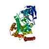
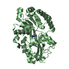
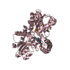
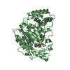
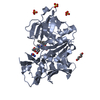
| Title | CRYSTAL STRUCTURE OF MANGANESE PEROXIDASE 4 FROM PLEUROTUS OSTREATUS - CRYSTAL FORM I | |||||||||
 Components Components | MANGANESE PEROXIDASE 4 | |||||||||
 Keywords Keywords | OXIDOREDUCTASE / CLASS II (FUNGAL) PEROXIDASES / PROTOPORPHYRIN IX / ELECTRON T LIGNIN PEROXIDASE / LIGNIN DEGRADATION | |||||||||
| Function / homology |  Function and homology information Function and homology informationOxidoreductases; Acting on a peroxide as acceptor; Peroxidases / hydrogen peroxide catabolic process / response to reactive oxygen species / peroxidase activity / cellular response to oxidative stress / heme binding / extracellular region / metal ion binding Similarity search - Function | |||||||||
| Biological species |  PLEUROTUS OSTREATUS (oyster mushroom) PLEUROTUS OSTREATUS (oyster mushroom) | |||||||||
| Method |  X-RAY DIFFRACTION / SYNCHROTRON / MOLECULAR REPLACEMENT / Resolution: 1.098 Å X-RAY DIFFRACTION / SYNCHROTRON / MOLECULAR REPLACEMENT / Resolution: 1.098 Å | |||||||||
 Authors Authors | Medrano, F.J. / Romero, A. | |||||||||
 Citation Citation | Journal: Biotechnol.Biofuels / Year: 2014 Title: Ligninolytic Peroxidase Genes in the Oyster Mushroom Genome: Heterologous Expression, Molecular Structure, Catalytic and Stability Properties, and Lignin-Degrading Ability. Authors: Fernandez-Fueyo, E. / Ruiz-Duenas, F.J. / Martinez, M.J. / Romero, A. / Hammel, K.E. / Medrano, F.J. / Martinez, A.T. | |||||||||
| History |
|
- Structure visualization
| Structure viewer | Molecule: MolmilJmol/JSmol |
|---|
- Downloads & links
-Download
| PDBx/mmCIF format | 4bm1.cif.gz | 396.7 KB | Display | PDBx/mmCIF format |
|---|---|---|---|---|
| PDB format | pdb4bm1.ent.gz | 328.9 KB | Display | PDB format |
| PDBx/mmJSON format | 4bm1.json.gz | Tree view | PDBx/mmJSON format | |
| Others |  Other downloads Other downloads |
-Validation report
| Arichive directory | https://data.pdbj.org/pub/pdb/validation_reports/bm/4bm1ftp://data.pdbj.org/pub/pdb/validation_reports/bm/4bm1 | HTTPS FTP |
|---|
-Related structure data
| Related structure data |  4blkC  4bllC  4blnC  4blxC  4blyC  4blzC  4bm0C 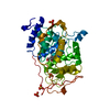 4bm2C 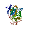 4bm3C 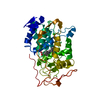 4bm4C  3fmuS C: citing same article ( S: Starting model for refinement |
|---|---|
| Similar structure data |


-Links
 PDBj
PDBj
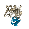
- Assembly
| Deposited unit | 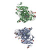
| ||||||||
|---|---|---|---|---|---|---|---|---|---|
| 1 | 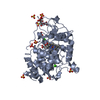
| ||||||||
| 2 | 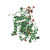
| ||||||||
| Unit cell |
|
-Components
-Protein , 1 types, 2 molecules AB
| #1: Protein | Mass: 36350.035 Da / Num. of mol.: 2 Source method: isolated from a genetically manipulated source Source: (gene. exp.) PLEUROTUS OSTREATUS (oyster mushroom) / Production host:  |
|---|
-Non-polymers , 5 types, 1236 molecules 


| #2: Chemical | ChemComp-CA /  Mass: 40.078 Da / Num. of mol.: 4 / Source method: obtained synthetically / Formula: Ca Mass: 40.078 Da / Num. of mol.: 4 / Source method: obtained synthetically / Formula: Ca#3: Chemical |  Mass: 616.487 Da / Num. of mol.: 2 / Source method: obtained synthetically / Formula: C34H32FeN4O4 Mass: 616.487 Da / Num. of mol.: 2 / Source method: obtained synthetically / Formula: C34H32FeN4O4#4: Chemical | ChemComp-SO4 /  Mass: 96.063 Da / Num. of mol.: 21 / Source method: obtained synthetically / Formula: SO4 Mass: 96.063 Da / Num. of mol.: 21 / Source method: obtained synthetically / Formula: SO4#5: Chemical |  Mass: 192.124 Da / Num. of mol.: 2 / Source method: obtained synthetically / Formula: C6H8O7 Mass: 192.124 Da / Num. of mol.: 2 / Source method: obtained synthetically / Formula: C6H8O7#6: Water | ChemComp-HOH / | Mass: 18.015 Da / Num. of mol.: 1207 / Source method: isolated from a natural source / Formula: H2O |
|---|
-Details
| Has protein modification | Y |
|---|
-Experimental details
-Experiment
| Experiment | Method: X-RAY DIFFRACTION / Number of used crystals: 1 |
|---|
- Sample preparation
| Crystal | Density Matthews: 2.68 Å3/Da / Density % sol: 54.16 % / Description: NONE |
|---|---|
| Crystal grow | Details: 0.1 M NA-CITRATE AT PH 5.5 & 2.0 M (NH4)2SO4. |
-Data collection
| Diffraction | Mean temperature: 100 K |
|---|---|
| Diffraction source | Source: SYNCHROTRON / Site: SLS  / Beamline: X06DA / Wavelength: 0.891976 / Beamline: X06DA / Wavelength: 0.891976 |
| Detector | Type: DECTRIS PILATUS / Detector: PIXEL / Date: Nov 6, 2012 |
| Radiation | Protocol: SINGLE WAVELENGTH / Monochromatic (M) / Laue (L): M / Scattering type: x-ray |
| Radiation wavelength | Wavelength: 0.891976 Å / Relative weight: 1 |
| Reflection | Resolution: 1.1→50 Å / Num. obs: 271193 / % possible obs: 83.9 % / Observed criterion σ(I): 2 / Redundancy: 13.5 % / Biso Wilson estimate: 8.92 Å2 / Rmerge(I) obs: 0.05 / Net I/σ(I): 13.5 |
| Reflection shell | Resolution: 1.1→1.16 Å / Redundancy: 1.9 % / Rmerge(I) obs: 0.54 / Mean I/σ(I) obs: 1.9 / % possible all: 43 |
- Processing
| Software |
| |||||||||||||||||||||||||||||||||||||||||||||||||||||||||||||||||||||||||||||||||||||||||||||||||||||||||||||||||||||||||||||||||||||||||||||||||||||||||||||||||||||||||||||||||||||||||||||||||||||||||||||||||||||||||
|---|---|---|---|---|---|---|---|---|---|---|---|---|---|---|---|---|---|---|---|---|---|---|---|---|---|---|---|---|---|---|---|---|---|---|---|---|---|---|---|---|---|---|---|---|---|---|---|---|---|---|---|---|---|---|---|---|---|---|---|---|---|---|---|---|---|---|---|---|---|---|---|---|---|---|---|---|---|---|---|---|---|---|---|---|---|---|---|---|---|---|---|---|---|---|---|---|---|---|---|---|---|---|---|---|---|---|---|---|---|---|---|---|---|---|---|---|---|---|---|---|---|---|---|---|---|---|---|---|---|---|---|---|---|---|---|---|---|---|---|---|---|---|---|---|---|---|---|---|---|---|---|---|---|---|---|---|---|---|---|---|---|---|---|---|---|---|---|---|---|---|---|---|---|---|---|---|---|---|---|---|---|---|---|---|---|---|---|---|---|---|---|---|---|---|---|---|---|---|---|---|---|---|---|---|---|---|---|---|---|---|---|---|---|---|---|---|---|---|
| Refinement | Method to determine structure: MOLECULAR REPLACEMENT Starting model: PDB ENTRY 3FMU Resolution: 1.098→38.148 Å / SU ML: 0.09 / σ(F): 1.96 / Phase error: 13.88 / Stereochemistry target values: ML
| |||||||||||||||||||||||||||||||||||||||||||||||||||||||||||||||||||||||||||||||||||||||||||||||||||||||||||||||||||||||||||||||||||||||||||||||||||||||||||||||||||||||||||||||||||||||||||||||||||||||||||||||||||||||||
| Solvent computation | Shrinkage radii: 0.6 Å / VDW probe radii: 0.9 Å / Solvent model: FLAT BULK SOLVENT MODEL / Bsol: 46.371 Å2 / ksol: 0.4 e/Å3 | |||||||||||||||||||||||||||||||||||||||||||||||||||||||||||||||||||||||||||||||||||||||||||||||||||||||||||||||||||||||||||||||||||||||||||||||||||||||||||||||||||||||||||||||||||||||||||||||||||||||||||||||||||||||||
| Displacement parameters |
| |||||||||||||||||||||||||||||||||||||||||||||||||||||||||||||||||||||||||||||||||||||||||||||||||||||||||||||||||||||||||||||||||||||||||||||||||||||||||||||||||||||||||||||||||||||||||||||||||||||||||||||||||||||||||
| Refinement step | Cycle: LAST / Resolution: 1.098→38.148 Å
| |||||||||||||||||||||||||||||||||||||||||||||||||||||||||||||||||||||||||||||||||||||||||||||||||||||||||||||||||||||||||||||||||||||||||||||||||||||||||||||||||||||||||||||||||||||||||||||||||||||||||||||||||||||||||
| Refine LS restraints |
| |||||||||||||||||||||||||||||||||||||||||||||||||||||||||||||||||||||||||||||||||||||||||||||||||||||||||||||||||||||||||||||||||||||||||||||||||||||||||||||||||||||||||||||||||||||||||||||||||||||||||||||||||||||||||
| LS refinement shell |
|
