 Movie
Movie Controller

[English] 日本語
 Yorodumi
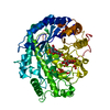
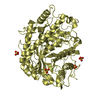
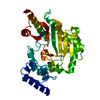
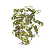
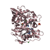
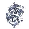
- PDB-4bhl: Crystal structure of Litopenaeus vannamei arginine kinase in bina... -
+ Open data

- Basic information
| Entry | Database: PDB / ID: 4bhl | ||||||
|---|---|---|---|---|---|---|---|
| Title | Crystal structure of Litopenaeus vannamei arginine kinase in binary complex with arginine | ||||||
 Components Components | ARGININE KINASE | ||||||
 Keywords Keywords | TRANSFERASE / BINARY COMPLEX / PHOSPHAGEN | ||||||
| Function / homology |  Function and homology information Function and homology informationdTDP metabolic process / arginine kinase / arginine kinase activity / phosphocreatine biosynthetic process / creatine kinase activity / arginine binding / : / ATP binding Similarity search - Function | ||||||
| Biological species |  LITOPENAEUS VANNAMEI (Pacific white shrimp) LITOPENAEUS VANNAMEI (Pacific white shrimp) | ||||||
| Method |  X-RAY DIFFRACTION / SYNCHROTRON / MOLECULAR REPLACEMENT / Resolution: 1.9 Å X-RAY DIFFRACTION / SYNCHROTRON / MOLECULAR REPLACEMENT / Resolution: 1.9 Å | ||||||
 Authors Authors | Lopez-Zavala, A.A. / Garcia-Orozco, K.D. / Carrasco-Miranda, J.S. / Hernandez-Flores, J.M. / Sugich-Miranda, R. / Velazquez-Contreras, E.F. / Criscitiello, M.F. / Brieba, L.G. / Rudino-Pinera, E. / Sotelo-Mundo, R.R. | ||||||
 Citation Citation | Journal: J.Bioenerg.Biomembr. / Year: 2013 Title: Crystal Structure of Shrimp Arginine Kinase in Binary Complex with Arginine-A Molecular View of the Phosphagen Precursor Binding to the Enzyme. Authors: Lopez-Zavala, A.A. / Garcia-Orozco, K.D. / Carrasco-Miranda, J.S. / Sugich-Miranda, R. / Velazquez-Contreras, E.F. / Criscitiello, M.F. / Brieba, L.G. / Rudino-Pinera, E. / Sotelo-Mundo, R.R. | ||||||
| History |
|
- Structure visualization
| Structure viewer | Molecule: MolmilJmol/JSmol |
|---|
- Downloads & links
-Download
| PDBx/mmCIF format | 4bhl.cif.gz | 88.4 KB | Display | PDBx/mmCIF format |
|---|---|---|---|---|
| PDB format | pdb4bhl.ent.gz | 64.6 KB | Display | PDB format |
| PDBx/mmJSON format | 4bhl.json.gz | Tree view | PDBx/mmJSON format | |
| Others |  Other downloads Other downloads |
-Validation report
| Arichive directory | https://data.pdbj.org/pub/pdb/validation_reports/bh/4bhlftp://data.pdbj.org/pub/pdb/validation_reports/bh/4bhl | HTTPS FTP |
|---|
-Related structure data
| Related structure data | 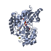 4bg4C 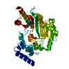 4am1S S: Starting model for refinement C: citing same article ( |
|---|---|
| Similar structure data |


-Links
 PDBj
PDBj


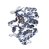
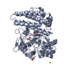
- Assembly
| Deposited unit | 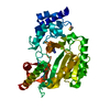
| ||||||||
|---|---|---|---|---|---|---|---|---|---|
| 1 |
| ||||||||
| Unit cell |
|
-Components
| #1: Protein | Mass: 40207.727 Da / Num. of mol.: 1 / Source method: isolated from a natural source Source: (natural) LITOPENAEUS VANNAMEI (Pacific white shrimp)Tissue: TAIL MUSCLE / References: UniProt: Q004B5, arginine kinase |
|---|---|
| #2: Chemical | ChemComp-ARG /   Type: L-peptide linking / Mass: 175.209 Da / Num. of mol.: 1 / Source method: obtained synthetically / Formula: C6H15N4O2 Type: L-peptide linking / Mass: 175.209 Da / Num. of mol.: 1 / Source method: obtained synthetically / Formula: C6H15N4O2 |
| #3: Chemical | ChemComp-BME /   Mass: 78.133 Da / Num. of mol.: 1 / Source method: obtained synthetically / Formula: C2H6OS Mass: 78.133 Da / Num. of mol.: 1 / Source method: obtained synthetically / Formula: C2H6OS |
| #4: Water | ChemComp-HOH /  Mass: 18.015 Da / Num. of mol.: 291 / Source method: isolated from a natural source / Formula: H2O Mass: 18.015 Da / Num. of mol.: 291 / Source method: isolated from a natural source / Formula: H2O |
| Nonpolymer details | BETA-MERCAPTOETHANOL (BME): BETA-MERCAPTOETHANOL IS COVALENTLY BOUNDED TO CYS 139. ARGININE (ARG): ...BETA-MERCAPTOET |
-Experimental details
-Experiment
| Experiment | Method: X-RAY DIFFRACTION / Number of used crystals: 1 |
|---|
- Sample preparation
| Crystal | Density Matthews: 2.03 Å3/Da / Density % sol: 39.5 % / Description: NONE |
|---|---|
| Crystal grow | Method: microbatch / pH: 6.5 Details: PROTEIN WAS CRYSTALLIZED FROM 0.2 M SODIUM ACETATE, 0.1 M SODIUM CACODYLATE PH 6.5 AND 30 % (W/V) PEG 8000 USING THE MICROBATCH METHOD |
-Data collection
| Diffraction | Mean temperature: 100 K |
|---|---|
| Diffraction source | Source: SYNCHROTRON / Site: NSLS  / Beamline: X6A / Wavelength: 0.9795 / Beamline: X6A / Wavelength: 0.9795 |
| Detector | Type: ADSC QUANTUM 270 / Detector: CCD / Date: Jul 5, 2012 Details: 1M LONG RH COATED TOROIDAL MIRROR FOR VERTICAL AND HORIZONTAL FOCUSING |
| Radiation | Monochromator: SI(111) CHANNEL CUT / Protocol: SINGLE WAVELENGTH / Monochromatic (M) / Laue (L): M / Scattering type: x-ray |
| Radiation wavelength | Wavelength: 0.9795 Å / Relative weight: 1 |
| Reflection | Resolution: 1.9→19.1 Å / Num. obs: 26516 / % possible obs: 100 % / Observed criterion σ(I): 3 / Redundancy: 7.1 % / Biso Wilson estimate: 14.29 Å2 / Rmerge(I) obs: 0.06 / Net I/σ(I): 22.3 |
| Reflection shell | Resolution: 1.9→2.01 Å / Redundancy: 6.9 % / Rmerge(I) obs: 0.21 / Mean I/σ(I) obs: 5.4 / % possible all: 96.1 |
- Processing
| Software |
| |||||||||||||||||||||||||||||||||||||||||||||||||||||||||||||||||||||||||||||
|---|---|---|---|---|---|---|---|---|---|---|---|---|---|---|---|---|---|---|---|---|---|---|---|---|---|---|---|---|---|---|---|---|---|---|---|---|---|---|---|---|---|---|---|---|---|---|---|---|---|---|---|---|---|---|---|---|---|---|---|---|---|---|---|---|---|---|---|---|---|---|---|---|---|---|---|---|---|---|
| Refinement | Method to determine structure: MOLECULAR REPLACEMENT Starting model: PDB ENTRY 4AM1 Resolution: 1.9→46.461 Å / SU ML: 0.2 / σ(F): 1.97 / Phase error: 18.62 / Stereochemistry target values: ML Details: RESIDUES 312-319 ARE DISORDERED AND WERE NOT INCLUDED IN THE FINAL MODEL.
| |||||||||||||||||||||||||||||||||||||||||||||||||||||||||||||||||||||||||||||
| Solvent computation | Shrinkage radii: 0.6 Å / VDW probe radii: 0.9 Å / Solvent model: FLAT BULK SOLVENT MODEL / Bsol: 51.96 Å2 / ksol: 0.412 e/Å3 | |||||||||||||||||||||||||||||||||||||||||||||||||||||||||||||||||||||||||||||
| Displacement parameters | Biso mean: 16.3 Å2
| |||||||||||||||||||||||||||||||||||||||||||||||||||||||||||||||||||||||||||||
| Refinement step | Cycle: LAST / Resolution: 1.9→46.461 Å
| |||||||||||||||||||||||||||||||||||||||||||||||||||||||||||||||||||||||||||||
| Refine LS restraints |
| |||||||||||||||||||||||||||||||||||||||||||||||||||||||||||||||||||||||||||||
| LS refinement shell |
|
