 Movie
Movie Controller

[English] 日本語
 Yorodumi
- PDB-1ukp: Crystal structure of soybean beta-amylase mutant substituted at s... -
+ Open data

- Basic information
| Entry | Database: PDB / ID: 1ukp | ||||||
|---|---|---|---|---|---|---|---|
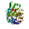
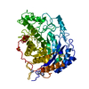
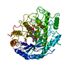
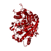
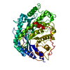
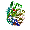
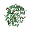
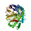
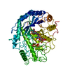
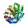
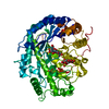
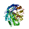
| Title | Crystal structure of soybean beta-amylase mutant substituted at surface region | ||||||
 Components Components | Beta-amylase | ||||||
 Keywords Keywords | HYDROLASE / (alpha/beta)8 barrel | ||||||
| Function / homology |  Function and homology information Function and homology information | ||||||
| Biological species |  | ||||||
| Method |  X-RAY DIFFRACTION / SYNCHROTRON / MOLECULAR REPLACEMENT / Resolution: 2.1 Å X-RAY DIFFRACTION / SYNCHROTRON / MOLECULAR REPLACEMENT / Resolution: 2.1 Å | ||||||
 Authors Authors | Kang, Y.N. / Adachi, M. / Mikami, B. / Utsumi, S. | ||||||
 Citation Citation | Journal: Protein Eng. / Year: 2003 Title: Change in the crystal packing of soybean beta-amylase mutants substituted at a few surface amino acid residues Authors: Kang, Y.N. / Adachi, M. / Mikami, B. / Utsumi, S. | ||||||
| History |
|
- Structure visualization
| Structure viewer | Molecule: MolmilJmol/JSmol |
|---|
- Downloads & links
-Download
| PDBx/mmCIF format | 1ukp.cif.gz | 407.6 KB | Display | PDBx/mmCIF format |
|---|---|---|---|---|
| PDB format | pdb1ukp.ent.gz | 332.8 KB | Display | PDB format |
| PDBx/mmJSON format | 1ukp.json.gz | Tree view | PDBx/mmJSON format | |
| Others |  Other downloads Other downloads |
-Validation report
| Arichive directory | https://data.pdbj.org/pub/pdb/validation_reports/uk/1ukpftp://data.pdbj.org/pub/pdb/validation_reports/uk/1ukp | HTTPS FTP |
|---|
-Related structure data
| Related structure data | 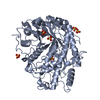 1ukoC 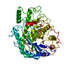 1bybS S: Starting model for refinement C: citing same article ( |
|---|---|
| Similar structure data |


-Links
 PDBj
PDBj- Assembly
| Deposited unit | 
| ||||||||
|---|---|---|---|---|---|---|---|---|---|
| 1 | 
| ||||||||
| 2 | 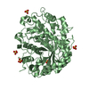
| ||||||||
| 3 | 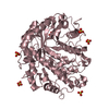
| ||||||||
| 4 | 
| ||||||||
| Unit cell |
| ||||||||
| Details | the biological assembly is a monomer |
-Components
| #1: Protein | Mass: 56033.168 Da / Num. of mol.: 4 / Mutation: D374Y / L481R / P487D / K462S Source method: isolated from a genetically manipulated source Source: (gene. exp.)  #2: Chemical | ChemComp-SO4 /   Mass: 96.063 Da / Num. of mol.: 20 / Source method: obtained synthetically / Formula: SO4 Mass: 96.063 Da / Num. of mol.: 20 / Source method: obtained synthetically / Formula: SO4#3: Water | ChemComp-HOH / |  Mass: 18.015 Da / Num. of mol.: 1005 / Source method: isolated from a natural source / Formula: H2O Mass: 18.015 Da / Num. of mol.: 1005 / Source method: isolated from a natural source / Formula: H2O |
|---|
-Experimental details
-Experiment
| Experiment | Method: X-RAY DIFFRACTION / Number of used crystals: 1 |
|---|
- Sample preparation
| Crystal | Density Matthews: 2.24 Å3/Da / Density % sol: 44.68 % | ||||||||||||||||||||||||||||||||||||
|---|---|---|---|---|---|---|---|---|---|---|---|---|---|---|---|---|---|---|---|---|---|---|---|---|---|---|---|---|---|---|---|---|---|---|---|---|---|
| Crystal grow | Temperature: 277 K / Method: vapor diffusion, hanging drop / pH: 5.4 Details: ammonium sulfate, sodium acetate, 2-mercaptoethanol, EDTA, pH 5.4, VAPOR DIFFUSION, HANGING DROP, temperature 277.0K | ||||||||||||||||||||||||||||||||||||
| Crystal grow | *PLUS Temperature: 4 ℃ / Method: vapor diffusion, hanging drop | ||||||||||||||||||||||||||||||||||||
| Components of the solutions | *PLUS
|
-Data collection
| Diffraction | Mean temperature: 100 K |
|---|---|
| Diffraction source | Source: SYNCHROTRON / Site: SPring-8  / Beamline: BL44XU / Wavelength: 0.9 Å / Beamline: BL44XU / Wavelength: 0.9 Å |
| Detector | Type: OXFORD PX210 / Detector: CCD / Date: Apr 17, 2002 |
| Radiation | Monochromator: rotating-inclining double crystal monochromator Protocol: SINGLE WAVELENGTH / Monochromatic (M) / Laue (L): M / Scattering type: x-ray |
| Radiation wavelength | Wavelength: 0.9 Å / Relative weight: 1 |
| Reflection | Resolution: 2→19.98 Å / Num. all: 335804 / Num. obs: 137751 / % possible obs: 95.4 % / Observed criterion σ(I): 1 / Redundancy: 2.44 % / Biso Wilson estimate: 16.2 Å2 / Rmerge(I) obs: 0.121 |
| Reflection shell | Resolution: 2→2.07 Å / Rmerge(I) obs: 0.383 / Num. unique all: 13776 / % possible all: 95.3 |
| Reflection | *PLUS Num. measured all: 335804 |
| Reflection shell | *PLUS % possible obs: 95.3 % / Num. unique obs: 13776 / Num. measured obs: 33459 |
- Processing
| Software |
| ||||||||||||||||||||||||||||||||||||
|---|---|---|---|---|---|---|---|---|---|---|---|---|---|---|---|---|---|---|---|---|---|---|---|---|---|---|---|---|---|---|---|---|---|---|---|---|---|
| Refinement | Method to determine structure: MOLECULAR REPLACEMENT Starting model: PDB ENTRY 1BYB Resolution: 2.1→10 Å / Rfactor Rfree error: 0.002 / Data cutoff high absF: 1432544.57 / Data cutoff low absF: 0 / Isotropic thermal model: RESTRAINED / Cross valid method: THROUGHOUT / σ(F): 0 / Stereochemistry target values: Engh & Huber
| ||||||||||||||||||||||||||||||||||||
| Solvent computation | Solvent model: FLAT MODEL / Bsol: 71.9499 Å2 / ksol: 0.455532 e/Å3 | ||||||||||||||||||||||||||||||||||||
| Displacement parameters | Biso mean: 24.1 Å2
| ||||||||||||||||||||||||||||||||||||
| Refine analyze |
| ||||||||||||||||||||||||||||||||||||
| Refinement step | Cycle: LAST / Resolution: 2.1→10 Å
| ||||||||||||||||||||||||||||||||||||
| Refine LS restraints |
| ||||||||||||||||||||||||||||||||||||
| Refine LS restraints NCS | NCS model details: CONSTR | ||||||||||||||||||||||||||||||||||||
| LS refinement shell | Resolution: 2.1→2.23 Å / Rfactor Rfree error: 0.006 / Total num. of bins used: 6
| ||||||||||||||||||||||||||||||||||||
| Xplor file |
| ||||||||||||||||||||||||||||||||||||
| Refinement | *PLUS Highest resolution: 2.1 Å / Lowest resolution: 10 Å / Rfactor Rfree: 0.2345 / Rfactor Rwork: 0.2001 | ||||||||||||||||||||||||||||||||||||
| Solvent computation | *PLUS | ||||||||||||||||||||||||||||||||||||
| Displacement parameters | *PLUS | ||||||||||||||||||||||||||||||||||||
| Refine LS restraints | *PLUS
| ||||||||||||||||||||||||||||||||||||
| LS refinement shell | *PLUS Lowest resolution: 2.17 Å / Num. reflection Rwork: 11208 |
