 Movie
Movie Controller

[English] 日本語
 Yorodumi
- PDB-2xgb: Crystal structure of Barley Beta-Amylase complexed with 2,3- epox... -
+ Open data

- Basic information
| Entry | Database: PDB / ID: 2xgb | ||||||
|---|---|---|---|---|---|---|---|
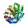
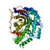
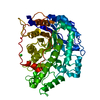
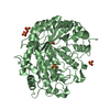
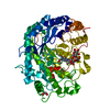
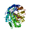
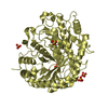
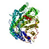
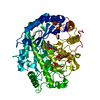
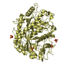
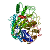
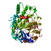
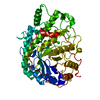
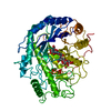
| Title | Crystal structure of Barley Beta-Amylase complexed with 2,3- epoxypropyl-alpha-D-glucopyranoside | ||||||
 Components Components | BETA-AMYLASE | ||||||
 Keywords Keywords | HYDROLASE / CARBOHYDRATE METABOLISM / GERMINATION | ||||||
| Function / homology |  Function and homology information Function and homology informationbeta-amylase / beta-amylase activity / polysaccharide catabolic process / identical protein binding Similarity search - Function | ||||||
| Biological species |  | ||||||
| Method |  X-RAY DIFFRACTION / SYNCHROTRON / MOLECULAR REPLACEMENT / Resolution: 1.2 Å X-RAY DIFFRACTION / SYNCHROTRON / MOLECULAR REPLACEMENT / Resolution: 1.2 Å | ||||||
 Authors Authors | Rejzek, M. / Stevenson, C.E.M. / Southard, A.M. / Stanley, D. / Denyer, K. / Smith, A.M. / Naldrett, M.J. / Lawson, D.M. / Field, R.A. | ||||||
 Citation Citation | Journal: Mol.Biosyst. / Year: 2011 Title: Chemical Genetics and Cereal Starch Metabolism: Structural Basis of the Non-Covalent and Covalent Inhibition of Barley Beta-Amylase. Authors: Rejzek, M. / Stevenson, C.E. / Southard, A.M. / Stanley, D. / Denyer, K. / Smith, A.M. / Naldrett, M.J. / Lawson, D.M. / Field, R.A. | ||||||
| History |
| ||||||
| Remark 700 | SHEET DETERMINATION METHOD: DSSP THE SHEETS PRESENTED AS "AA" IN EACH CHAIN ON SHEET RECORDS BELOW ... SHEET DETERMINATION METHOD: DSSP THE SHEETS PRESENTED AS "AA" IN EACH CHAIN ON SHEET RECORDS BELOW IS ACTUALLY AN 8-STRANDED BARREL THIS IS REPRESENTED BY A 9-STRANDED SHEET IN WHICH THE FIRST AND LAST STRANDS ARE IDENTICAL. |
- Structure visualization
| Structure viewer | Molecule: MolmilJmol/JSmol |
|---|
- Downloads & links
-Download
| PDBx/mmCIF format | 2xgb.cif.gz | 243.3 KB | Display | PDBx/mmCIF format |
|---|---|---|---|---|
| PDB format | pdb2xgb.ent.gz | 196.4 KB | Display | PDB format |
| PDBx/mmJSON format | 2xgb.json.gz | Tree view | PDBx/mmJSON format | |
| Others |  Other downloads Other downloads |
-Validation report
| Arichive directory | https://data.pdbj.org/pub/pdb/validation_reports/xg/2xgbftp://data.pdbj.org/pub/pdb/validation_reports/xg/2xgb | HTTPS FTP |
|---|
-Related structure data
| Related structure data |  2xffC 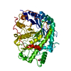 2xfrC 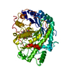 2xfyC 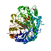 2xg9C 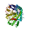 2xgiC 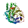 1b1yS C: citing same article ( S: Starting model for refinement |
|---|---|
| Similar structure data |


-Links
 PDBj
PDBj- Assembly
| Deposited unit | 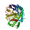
| ||||||||
|---|---|---|---|---|---|---|---|---|---|
| 1 |
| ||||||||
| Unit cell |
|
-Components
| #1: Protein | Mass: 59670.102 Da / Num. of mol.: 1 / Source method: isolated from a natural source / Details: PROTEIN PURCHASED FROM MEGAZYME / Source: (natural) | ||||||||
|---|---|---|---|---|---|---|---|---|---|
| #2: Sugar | ChemComp-EPG / (  Type: D-saccharide / Mass: 236.219 Da / Num. of mol.: 1 Type: D-saccharide / Mass: 236.219 Da / Num. of mol.: 1Source method: isolated from a genetically manipulated source Formula: C9H16O7 | ||||||||
| #3: Chemical | ChemComp-EDO /   Mass: 62.068 Da / Num. of mol.: 4 / Source method: obtained synthetically / Formula: C2H6O2 Mass: 62.068 Da / Num. of mol.: 4 / Source method: obtained synthetically / Formula: C2H6O2#4: Water | ChemComp-HOH / |  Mass: 18.015 Da / Num. of mol.: 729 / Source method: isolated from a natural source / Formula: H2O Mass: 18.015 Da / Num. of mol.: 729 / Source method: isolated from a natural source / Formula: H2OHas protein modification | Y | Nonpolymer details | 1,2-ETHANEDIOL (EDO): PRESENT AT 20 PERCENT IN THE CRYOPROTECTANT 2,3-EPOXYPROPYL-ALPHA-D- ...1,2-ETHANEDIOL | Sequence details | PROTEIN ISOLATED FROM NATURAL SOURCE BUT SEQUENCE DIFFERS AT 3 POSITIONS TO UNIPROTKB DATABASE ...PROTEIN ISOLATED FROM NATURAL SOURCE BUT SEQUENCE DIFFERS AT 3 POSITIONS TO UNIPROTKB DATABASE ENTRY P16098. THESE CHANGES WERE IDENTIFIED | |
-Experimental details
-Experiment
| Experiment | Method: X-RAY DIFFRACTION / Number of used crystals: 1 |
|---|
- Sample preparation
| Crystal | Density Matthews: 1.91 Å3/Da / Density % sol: 35.6 % / Description: NONE |
|---|---|
| Crystal grow | Temperature: 291 K / Method: vapor diffusion, hanging drop / pH: 5.5 Details: CRYSTALS WERE GROWN AT 291 K USING THE HANGING DROP VAPOUR DIFFUSION METHOD WITH PROTEIN AT 10 MG PER ML AND A PRECIPITANT COMPRISED OF 14 PERCENT PEG 3350 IN 100 MM BIS-TRIS PROPANE BUFFER AT PH 5.5 |
-Data collection
| Diffraction | Mean temperature: 100 K |
|---|---|
| Diffraction source | Source: SYNCHROTRON / Site: SRS  / Beamline: PX10.1 / Wavelength: 1.117 / Beamline: PX10.1 / Wavelength: 1.117 |
| Detector | Type: MARMOSAIC 225 mm CCD / Detector: CCD / Date: Nov 12, 2007 |
| Radiation | Protocol: SINGLE WAVELENGTH / Monochromatic (M) / Laue (L): M / Scattering type: x-ray |
| Radiation wavelength | Wavelength: 1.117 Å / Relative weight: 1 |
| Reflection | Resolution: 1.11→33.69 Å / Num. obs: 135507 / % possible obs: 96.3 % / Observed criterion σ(I): -9 / Redundancy: 5.98 % / Biso Wilson estimate: 10.6 Å2 / Rmerge(I) obs: 0.1 / Net I/σ(I): 3.56 |
| Reflection shell | Resolution: 1.2→1.26 Å / Redundancy: 3.01 % / Rmerge(I) obs: 0.27 / Mean I/σ(I) obs: 2.76 / % possible all: 79.5 |
- Processing
| Software |
| ||||||||||||||||||||||||||||||||||||||||||||||||||||||||||||||||||||||||||||||||||||||||||||||||||||||||||||||||||||||||||||||||||||||||||||||||||||||||||||||||||||||||||||||||||||||
|---|---|---|---|---|---|---|---|---|---|---|---|---|---|---|---|---|---|---|---|---|---|---|---|---|---|---|---|---|---|---|---|---|---|---|---|---|---|---|---|---|---|---|---|---|---|---|---|---|---|---|---|---|---|---|---|---|---|---|---|---|---|---|---|---|---|---|---|---|---|---|---|---|---|---|---|---|---|---|---|---|---|---|---|---|---|---|---|---|---|---|---|---|---|---|---|---|---|---|---|---|---|---|---|---|---|---|---|---|---|---|---|---|---|---|---|---|---|---|---|---|---|---|---|---|---|---|---|---|---|---|---|---|---|---|---|---|---|---|---|---|---|---|---|---|---|---|---|---|---|---|---|---|---|---|---|---|---|---|---|---|---|---|---|---|---|---|---|---|---|---|---|---|---|---|---|---|---|---|---|---|---|---|---|
| Refinement | Method to determine structure: MOLECULAR REPLACEMENT Starting model: PDB ENTRY 1B1Y Resolution: 1.2→33.168 Å / Cor.coef. Fo:Fc: 0.981 / Cor.coef. Fo:Fc free: 0.977 / SU B: 0.833 / SU ML: 0.017 / Cross valid method: THROUGHOUT / σ(F): 0 / ESU R: 0.035 / ESU R Free: 0.034 / Stereochemistry target values: MAXIMUM LIKELIHOOD / Details: HYDROGENS HAVE BEEN ADDED IN THE RIDING POSITIONS.
| ||||||||||||||||||||||||||||||||||||||||||||||||||||||||||||||||||||||||||||||||||||||||||||||||||||||||||||||||||||||||||||||||||||||||||||||||||||||||||||||||||||||||||||||||||||||
| Solvent computation | Ion probe radii: 0.8 Å / Shrinkage radii: 0.8 Å / VDW probe radii: 1.4 Å / Solvent model: MASK BULK SOLVENT | ||||||||||||||||||||||||||||||||||||||||||||||||||||||||||||||||||||||||||||||||||||||||||||||||||||||||||||||||||||||||||||||||||||||||||||||||||||||||||||||||||||||||||||||||||||||
| Displacement parameters | Biso mean: 9.8 Å2
| ||||||||||||||||||||||||||||||||||||||||||||||||||||||||||||||||||||||||||||||||||||||||||||||||||||||||||||||||||||||||||||||||||||||||||||||||||||||||||||||||||||||||||||||||||||||
| Refinement step | Cycle: LAST / Resolution: 1.2→33.168 Å
| ||||||||||||||||||||||||||||||||||||||||||||||||||||||||||||||||||||||||||||||||||||||||||||||||||||||||||||||||||||||||||||||||||||||||||||||||||||||||||||||||||||||||||||||||||||||
| Refine LS restraints |
|