 Movie
Movie Controller

[English] 日本語
 Yorodumi
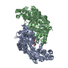
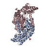
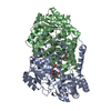
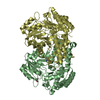
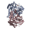
- PDB-4bhd: Methanococcus jannaschii serine hydroxymethyl-transferase, apo form -
+ Open data

- Basic information
| Entry | Database: PDB / ID: 4bhd | ||||||
|---|---|---|---|---|---|---|---|
| Title | Methanococcus jannaschii serine hydroxymethyl-transferase, apo form | ||||||
 Components Components | SERINE HYDROXYMETHYLTRANSFERASE | ||||||
 Keywords Keywords | TRANSFERASE | ||||||
| Function / homology |  Function and homology information Function and homology informationTransferases; Transferring one-carbon groups; Hydroxymethyl-, formyl- and related transferases / L-allo-threonine aldolase / L-allo-threonine aldolase activity / glycine hydroxymethyltransferase activity / : / tetrahydrofolate metabolic process / tetrahydrofolate interconversion / pyridoxal phosphate binding / cytoplasm Similarity search - Function | ||||||
| Biological species |   METHANOCALDOCOCCUS JANNASCHII (archaea) METHANOCALDOCOCCUS JANNASCHII (archaea) | ||||||
| Method |  X-RAY DIFFRACTION / SYNCHROTRON / MOLECULAR REPLACEMENT / Resolution: 2.83 Å X-RAY DIFFRACTION / SYNCHROTRON / MOLECULAR REPLACEMENT / Resolution: 2.83 Å | ||||||
 Authors Authors | Saccoccia, F. / Angelucci, F. / Ilari, A. | ||||||
 Citation Citation | Journal: Proteins / Year: 2014 Title: The Crystal Structure of Archaeal Serine Hydroxymethyltransferase Reveals Idiosyncratic Features Likely Required to Withstand High Temperatures. Authors: Angelucci, F. / Morea, V. / Angelaccio, S. / Saccoccia, F. / Contestabile, R. / Ilari, A. | ||||||
| History |
|
- Structure visualization
| Structure viewer | Molecule: MolmilJmol/JSmol |
|---|
- Downloads & links
-Download
| PDBx/mmCIF format | 4bhd.cif.gz | 332 KB | Display | PDBx/mmCIF format |
|---|---|---|---|---|
| PDB format | pdb4bhd.ent.gz | 274.4 KB | Display | PDB format |
| PDBx/mmJSON format | 4bhd.json.gz | Tree view | PDBx/mmJSON format | |
| Others |  Other downloads Other downloads |
-Validation report
| Arichive directory | https://data.pdbj.org/pub/pdb/validation_reports/bh/4bhdftp://data.pdbj.org/pub/pdb/validation_reports/bh/4bhd | HTTPS FTP |
|---|
-Related structure data
| Related structure data |  4uqvC  1kl2S C: citing same article ( S: Starting model for refinement |
|---|---|
| Similar structure data |


-Links
 PDBj
PDBj- Assembly
| Deposited unit | 
| |||||||||||||||||||||||||||||||||||||||||||||||||||||||||||
|---|---|---|---|---|---|---|---|---|---|---|---|---|---|---|---|---|---|---|---|---|---|---|---|---|---|---|---|---|---|---|---|---|---|---|---|---|---|---|---|---|---|---|---|---|---|---|---|---|---|---|---|---|---|---|---|---|---|---|---|---|
| 1 |
| |||||||||||||||||||||||||||||||||||||||||||||||||||||||||||
| Unit cell |
| |||||||||||||||||||||||||||||||||||||||||||||||||||||||||||
| Noncrystallographic symmetry (NCS) | NCS domain:
NCS domain segments: Ens-ID: 1 / Refine code: 5
|
-Components
| #1: Protein | Mass: 48020.645 Da / Num. of mol.: 2 Source method: isolated from a genetically manipulated source Source: (gene. exp.) METHANOCALDOCOCCUS JANNASCHII (archaea)Production host:  References: UniProt: Q58992, glycine hydroxymethyltransferase, L-allo-threonine aldolase #2: Chemical | ChemComp-PO4 / |   Mass: 94.971 Da / Num. of mol.: 1 / Source method: obtained synthetically / Formula: PO4 Mass: 94.971 Da / Num. of mol.: 1 / Source method: obtained synthetically / Formula: PO4#3: Water | ChemComp-HOH / |  Mass: 18.015 Da / Num. of mol.: 16 / Source method: isolated from a natural source / Formula: H2O Mass: 18.015 Da / Num. of mol.: 16 / Source method: isolated from a natural source / Formula: H2O |
|---|
-Experimental details
-Experiment
| Experiment | Method: X-RAY DIFFRACTION / Number of used crystals: 1 |
|---|
- Sample preparation
| Crystal | Density Matthews: 2.74 Å3/Da / Density % sol: 55.22 % / Description: NONE |
|---|---|
| Crystal grow | pH: 5 / Details: NAH2PO4/K2HPO4 1.8 M, PH 5.0 |
-Data collection
| Diffraction | Mean temperature: 100 K |
|---|---|
| Diffraction source | Source: SYNCHROTRON / Site: ESRF  / Beamline: ID23-1 / Wavelength: 0.972 / Beamline: ID23-1 / Wavelength: 0.972 |
| Detector | Type: ADSC QUANTUM 315r / Detector: CCD |
| Radiation | Protocol: SINGLE WAVELENGTH / Monochromatic (M) / Laue (L): M / Scattering type: x-ray |
| Radiation wavelength | Wavelength: 0.972 Å / Relative weight: 1 |
| Reflection | Resolution: 2.85→50 Å / Num. obs: 25528 / % possible obs: 98.5 % / Observed criterion σ(I): -3 / Redundancy: 5.9 % / Rmerge(I) obs: 0.08 / Net I/σ(I): 13.93 |
| Reflection shell | Resolution: 2.85→2.95 Å / Redundancy: 5.5 % / Rmerge(I) obs: 0.5 / Mean I/σ(I) obs: 2.53 / % possible all: 98.9 |
- Processing
| Software |
| ||||||||||||||||||||||||||||||||||||||||||||||||||||||||||||||||||||||||||||||||||||||||||||||||||||||||||||||||||||||||||||||||||||||||||||||||||||||||||||||||||||||||||||||||||||||
|---|---|---|---|---|---|---|---|---|---|---|---|---|---|---|---|---|---|---|---|---|---|---|---|---|---|---|---|---|---|---|---|---|---|---|---|---|---|---|---|---|---|---|---|---|---|---|---|---|---|---|---|---|---|---|---|---|---|---|---|---|---|---|---|---|---|---|---|---|---|---|---|---|---|---|---|---|---|---|---|---|---|---|---|---|---|---|---|---|---|---|---|---|---|---|---|---|---|---|---|---|---|---|---|---|---|---|---|---|---|---|---|---|---|---|---|---|---|---|---|---|---|---|---|---|---|---|---|---|---|---|---|---|---|---|---|---|---|---|---|---|---|---|---|---|---|---|---|---|---|---|---|---|---|---|---|---|---|---|---|---|---|---|---|---|---|---|---|---|---|---|---|---|---|---|---|---|---|---|---|---|---|---|---|
| Refinement | Method to determine structure: MOLECULAR REPLACEMENT Starting model: PDB ENTRY 1KL2 Resolution: 2.83→50 Å / Cor.coef. Fo:Fc: 0.918 / Cor.coef. Fo:Fc free: 0.865 / SU B: 40.373 / SU ML: 0.356 / Cross valid method: THROUGHOUT / ESU R Free: 0.444 / Stereochemistry target values: MAXIMUM LIKELIHOOD / Details: HYDROGENS HAVE BEEN ADDED IN THE RIDING POSITIONS.
| ||||||||||||||||||||||||||||||||||||||||||||||||||||||||||||||||||||||||||||||||||||||||||||||||||||||||||||||||||||||||||||||||||||||||||||||||||||||||||||||||||||||||||||||||||||||
| Solvent computation | Ion probe radii: 0.8 Å / Shrinkage radii: 0.8 Å / VDW probe radii: 1.4 Å / Solvent model: MASK | ||||||||||||||||||||||||||||||||||||||||||||||||||||||||||||||||||||||||||||||||||||||||||||||||||||||||||||||||||||||||||||||||||||||||||||||||||||||||||||||||||||||||||||||||||||||
| Displacement parameters | Biso mean: 85.509 Å2
| ||||||||||||||||||||||||||||||||||||||||||||||||||||||||||||||||||||||||||||||||||||||||||||||||||||||||||||||||||||||||||||||||||||||||||||||||||||||||||||||||||||||||||||||||||||||
| Refinement step | Cycle: LAST / Resolution: 2.83→50 Å
| ||||||||||||||||||||||||||||||||||||||||||||||||||||||||||||||||||||||||||||||||||||||||||||||||||||||||||||||||||||||||||||||||||||||||||||||||||||||||||||||||||||||||||||||||||||||
| Refine LS restraints |
|