 Movie
Movie Controller

[English] 日本語
 Yorodumi
- PDB-6ti3: Apo-SHMT from Streptococcus thermophilus Tyr55Ser variant in comp... -
+ Open data

- Basic information
| Entry | Database: PDB / ID: 6ti3 | ||||||
|---|---|---|---|---|---|---|---|
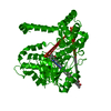
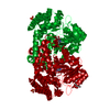
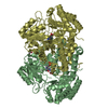
| Title | Apo-SHMT from Streptococcus thermophilus Tyr55Ser variant in complex with D-Threonine | ||||||
 Components Components | Serine hydroxymethyltransferase | ||||||
 Keywords Keywords | TRANSFERASE / Pyridoxal phosphate / X-ray crystallography / hydroxymethyltransferase / proton abstraction / tetrahydrofolate-independent | ||||||
| Function / homology |  Function and homology information Function and homology informationglycine hydroxymethyltransferase / glycine hydroxymethyltransferase activity / : / tetrahydrofolate interconversion / pyridoxal phosphate binding / cytosol Similarity search - Function | ||||||
| Biological species |  Streptococcus thermophilus (bacteria) Streptococcus thermophilus (bacteria) | ||||||
| Method |  X-RAY DIFFRACTION / SYNCHROTRON / MOLECULAR REPLACEMENT / Resolution: 1.96 Å X-RAY DIFFRACTION / SYNCHROTRON / MOLECULAR REPLACEMENT / Resolution: 1.96 Å | ||||||
 Authors Authors | Petrillo, G. / Hernandez, K. / Bujons, J. / Clapes, P. / Uson, I. | ||||||
| Funding support |  Spain, 1items Spain, 1items
| ||||||
 Citation Citation | Journal: To Be Published Title: Structural insights into nucleophile substrate specificity in variants of N-Serine hydroxymethyltransferase from Streptococcus thermophilus Authors: Petrillo, G. / Hernandez, K. / Bujons, J. / Clapes, P. / Uson, I. | ||||||
| History |
|
- Structure visualization
| Structure viewer | Molecule: MolmilJmol/JSmol |
|---|
- Downloads & links
-Download
| PDBx/mmCIF format | 6ti3.cif.gz | 333.1 KB | Display | PDBx/mmCIF format |
|---|---|---|---|---|
| PDB format | pdb6ti3.ent.gz | 271.3 KB | Display | PDB format |
| PDBx/mmJSON format | 6ti3.json.gz | Tree view | PDBx/mmJSON format | |
| Others |  Other downloads Other downloads |
-Validation report
| Arichive directory | https://data.pdbj.org/pub/pdb/validation_reports/ti/6ti3ftp://data.pdbj.org/pub/pdb/validation_reports/ti/6ti3 | HTTPS FTP |
|---|
-Related structure data
| Related structure data |  6tghC  6ti1C  6yrwC  4wxgS S: Starting model for refinement C: citing same article ( |
|---|---|
| Similar structure data |


-Links
 PDBj
PDBj- Assembly
| Deposited unit | 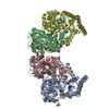
| ||||||||
|---|---|---|---|---|---|---|---|---|---|
| 1 | 
| ||||||||
| 2 | 
| ||||||||
| Unit cell |
|
-Components
| #1: Protein | Mass: 45061.926 Da / Num. of mol.: 4 Source method: isolated from a genetically manipulated source Source: (gene. exp.) Streptococcus thermophilus (bacteria) / Gene: glyA, CDA68_00974, DID82_07515 / Production host: References: UniProt: Q5MCK9, UniProt: Q5M0B4*PLUS, glycine hydroxymethyltransferase #2: Chemical |   Mass: 92.094 Da / Num. of mol.: 3 / Source method: isolated from a natural source / Formula: C3H8O3 / Feature type: SUBJECT OF INVESTIGATION Mass: 92.094 Da / Num. of mol.: 3 / Source method: isolated from a natural source / Formula: C3H8O3 / Feature type: SUBJECT OF INVESTIGATION#3: Chemical | ChemComp-DTH /   Type: D-peptide linking / Mass: 119.119 Da / Num. of mol.: 4 / Source method: obtained synthetically / Formula: C4H9NO3 / Feature type: SUBJECT OF INVESTIGATION Type: D-peptide linking / Mass: 119.119 Da / Num. of mol.: 4 / Source method: obtained synthetically / Formula: C4H9NO3 / Feature type: SUBJECT OF INVESTIGATION#4: Chemical | ChemComp-NA / |   Mass: 22.990 Da / Num. of mol.: 1 / Source method: obtained synthetically / Formula: Na / Feature type: SUBJECT OF INVESTIGATION Mass: 22.990 Da / Num. of mol.: 1 / Source method: obtained synthetically / Formula: Na / Feature type: SUBJECT OF INVESTIGATION#5: Water | ChemComp-HOH / |  Mass: 18.015 Da / Num. of mol.: 825 / Source method: isolated from a natural source / Formula: H2O Mass: 18.015 Da / Num. of mol.: 825 / Source method: isolated from a natural source / Formula: H2OHas ligand of interest | Y | |
|---|
-Experimental details
-Experiment
| Experiment | Method: X-RAY DIFFRACTION / Number of used crystals: 1 |
|---|
- Sample preparation
| Crystal | Density Matthews: 4.27 Å3/Da / Density % sol: 71.17 % |
|---|---|
| Crystal grow | Temperature: 293.15 K / Method: vapor diffusion, sitting drop Details: PLP 0.1M D-threonine 5mM Cacodylate 0.1M ph 6.5 Sodium citrate 1.2M |
-Data collection
| Diffraction | Mean temperature: 100 K / Serial crystal experiment: N |
|---|---|
| Diffraction source | Source: SYNCHROTRON / Site: ALBA / Beamline: XALOC / Wavelength: 0.933 Å |
| Detector | Type: DECTRIS PILATUS 6M / Detector: PIXEL / Date: Jul 20, 2016 |
| Radiation | Protocol: SINGLE WAVELENGTH / Monochromatic (M) / Laue (L): M / Scattering type: x-ray |
| Radiation wavelength | Wavelength: 0.933 Å / Relative weight: 1 |
| Reflection | Resolution: 1.96→100.09 Å / Num. obs: 208923 / % possible obs: 98.78 % / Redundancy: 8.1 % / Rmerge(I) obs: 0.09 / Net I/σ(I): 9.8 |
| Reflection shell | Resolution: 1.965→2.016 Å / Rmerge(I) obs: 0.09 / Num. unique obs: 15299 |
- Processing
| Software |
| ||||||||||||||||||||||||||||||||||||||||||||||||||||||||||||
|---|---|---|---|---|---|---|---|---|---|---|---|---|---|---|---|---|---|---|---|---|---|---|---|---|---|---|---|---|---|---|---|---|---|---|---|---|---|---|---|---|---|---|---|---|---|---|---|---|---|---|---|---|---|---|---|---|---|---|---|---|---|
| Refinement | Method to determine structure: MOLECULAR REPLACEMENT Starting model: 4WXG Resolution: 1.96→100.09 Å / Cor.coef. Fo:Fc: 0.968 / SU B: 2.792 / SU ML: 0.074 / Cross valid method: THROUGHOUT / σ(F): 0 / ESU R: 0.104 Details: HYDROGENS HAVE BEEN ADDED IN THE RIDING POSITIONS U VALUES : REFINED INDIVIDUALLY
| ||||||||||||||||||||||||||||||||||||||||||||||||||||||||||||
| Solvent computation | Ion probe radii: 0.8 Å / Shrinkage radii: 0.8 Å / VDW probe radii: 1.2 Å | ||||||||||||||||||||||||||||||||||||||||||||||||||||||||||||
| Displacement parameters | Biso max: 127.81 Å2 / Biso mean: 42.781 Å2 / Biso min: 20.35 Å2
| ||||||||||||||||||||||||||||||||||||||||||||||||||||||||||||
| Refinement step | Cycle: final / Resolution: 1.96→100.09 Å
| ||||||||||||||||||||||||||||||||||||||||||||||||||||||||||||
| Refine LS restraints |
| ||||||||||||||||||||||||||||||||||||||||||||||||||||||||||||
| LS refinement shell | Resolution: 1.965→2.016 Å / Rfactor Rfree error: 0 / Total num. of bins used: 20
|
