 Movie
Movie Controller


+ Open data

- Basic information
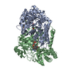
| Entry | Database: PDB / ID: 5yg4 | ||||||
|---|---|---|---|---|---|---|---|
| Title | Plasmodium vivax SHMT bound with PLP-glycine and S-GS849 | ||||||
 Components Components | Serine hydroxymethyltransferase | ||||||
 Keywords Keywords | TRANSFERASE/INHIBITOR / alpha and beta protein / Transferase / methyltransferase activity / Inhibitor / TRANSFERASE-INHIBITOR complex | ||||||
| Function / homology |  Function and homology information Function and homology informationglycine hydroxymethyltransferase / glycine hydroxymethyltransferase activity / : / tetrahydrofolate interconversion / methyltransferase activity / pyridoxal phosphate binding / methylation / mitochondrion Similarity search - Function | ||||||
| Biological species |  | ||||||
| Method |  X-RAY DIFFRACTION / MOLECULAR REPLACEMENT / Resolution: 2.3 Å X-RAY DIFFRACTION / MOLECULAR REPLACEMENT / Resolution: 2.3 Å | ||||||
| Model details | Plasmodium vivax SHMT bound with PLP-glycine and GS834 | ||||||
 Authors Authors | Chitnumsub, P. / Jaruwat, A. / Leartsakulpanich, U. / Schwertz, G. / Diederich, F. | ||||||
 Citation Citation | Journal: ChemMedChem / Year: 2018 Title: Potent Inhibitors of Plasmodial Serine Hydroxymethyltransferase (SHMT) Featuring a Spirocyclic Scaffold Authors: Schwertz, G. / Witschel, M.C. / Rottmann, M. / Leartsakulpanich, U. / Chitnumsub, P. / Jaruwat, A. / Amornwatcharapong, W. / Ittarat, W. / Schafer, A. / Aponte, R.A. / Trapp, N. / Chaiyen, P. / Diederich, F. | ||||||
| History |
|
- Structure visualization
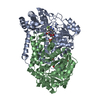
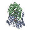
| Structure viewer | Molecule: MolmilJmol/JSmol |
|---|
- Downloads & links
-Download
| PDBx/mmCIF format | 5yg4.cif.gz | 275.1 KB | Display | PDBx/mmCIF format |
|---|---|---|---|---|
| PDB format | pdb5yg4.ent.gz | 222.8 KB | Display | PDB format |
| PDBx/mmJSON format | 5yg4.json.gz | Tree view | PDBx/mmJSON format | |
| Others |  Other downloads Other downloads |
-Validation report
| Arichive directory | https://data.pdbj.org/pub/pdb/validation_reports/yg/5yg4ftp://data.pdbj.org/pub/pdb/validation_reports/yg/5yg4 | HTTPS FTP |
|---|
-Related structure data
| Related structure data |  5yfzC  5yg2C  5yg3C  4tmrS S: Starting model for refinement C: citing same article ( |
|---|---|
| Similar structure data |


-Links
 PDBj
PDBj- Assembly
| Deposited unit | 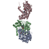
| ||||||||
|---|---|---|---|---|---|---|---|---|---|
| 1 | 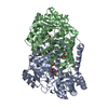
| ||||||||
| 2 | 
| ||||||||
| Unit cell |
| ||||||||
| Components on special symmetry positions |
|
-Components
-Protein , 1 types, 3 molecules ABC
| #1: Protein | Mass: 49249.160 Da / Num. of mol.: 3 Source method: isolated from a genetically manipulated source Source: (gene. exp.) Gene: PVC01_140059500, PVP01_1453700, PVT01_140059000 / Plasmid: pET17b / Production host:  References: UniProt: A0A1G4H5I1, glycine hydroxymethyltransferase |
|---|
-Non-polymers , 5 types, 381 molecules 


| #2: Chemical |  Mass: 306.209 Da / Num. of mol.: 3 / Source method: obtained synthetically / Formula: C10H15N2O7P Mass: 306.209 Da / Num. of mol.: 3 / Source method: obtained synthetically / Formula: C10H15N2O7P#3: Chemical |  Mass: 465.520 Da / Num. of mol.: 3 / Source method: obtained synthetically / Formula: C25H28FN5O3 Mass: 465.520 Da / Num. of mol.: 3 / Source method: obtained synthetically / Formula: C25H28FN5O3#4: Chemical |  Mass: 35.453 Da / Num. of mol.: 2 / Source method: obtained synthetically / Formula: Cl Mass: 35.453 Da / Num. of mol.: 2 / Source method: obtained synthetically / Formula: Cl#5: Chemical |  Mass: 92.094 Da / Num. of mol.: 3 / Source method: obtained synthetically / Formula: C3H8O3 Mass: 92.094 Da / Num. of mol.: 3 / Source method: obtained synthetically / Formula: C3H8O3#6: Water | ChemComp-HOH / | Mass: 18.015 Da / Num. of mol.: 370 / Source method: isolated from a natural source / Formula: H2O |
|---|
-Experimental details
-Experiment
| Experiment | Method: X-RAY DIFFRACTION / Number of used crystals: 1 |
|---|
- Sample preparation
| Crystal | Density Matthews: 2.33 Å3/Da / Density % sol: 47.19 % / Mosaicity: 0 ° |
|---|---|
| Crystal grow | Temperature: 298 K / Method: microbatch / pH: 8.5 / Details: PEG 4000, 0.06-0.12M NaCl, 0.1M Tris-HCl pH 8.5 |
-Data collection
| Diffraction | Mean temperature: 100 K | ||||||||||||||||||||||||
|---|---|---|---|---|---|---|---|---|---|---|---|---|---|---|---|---|---|---|---|---|---|---|---|---|---|
| Diffraction source | Source: ROTATING ANODE / Type: BRUKER D8 QUEST / Wavelength: 1.54 Å | ||||||||||||||||||||||||
| Detector | Type: BRUKER PHOTON 100 / Detector: CMOS / Date: Jul 21, 2017 | ||||||||||||||||||||||||
| Radiation | Protocol: SINGLE WAVELENGTH / Monochromatic (M) / Laue (L): M / Scattering type: x-ray | ||||||||||||||||||||||||
| Radiation wavelength | Wavelength: 1.54 Å / Relative weight: 1 | ||||||||||||||||||||||||
| Reflection | Resolution: 2.3→30 Å / Num. obs: 60621 / % possible obs: 99.5 % / Redundancy: 6.3 % / CC1/2: 0.998 / Rmerge(I) obs: 0.071 / Rpim(I) all: 0.029 / Rrim(I) all: 0.077 / Net I/σ(I): 17 | ||||||||||||||||||||||||
| Reflection shell | Diffraction-ID: 1
|
- Processing
| Software |
| ||||||||||||||||||||||||||||||||||||||||||||||||||||||||||||
|---|---|---|---|---|---|---|---|---|---|---|---|---|---|---|---|---|---|---|---|---|---|---|---|---|---|---|---|---|---|---|---|---|---|---|---|---|---|---|---|---|---|---|---|---|---|---|---|---|---|---|---|---|---|---|---|---|---|---|---|---|---|
| Refinement | Method to determine structure: MOLECULAR REPLACEMENT Starting model: 4TMR Resolution: 2.3→30 Å / Cor.coef. Fo:Fc: 0.925 / Cor.coef. Fo:Fc free: 0.875 / SU B: 10.188 / SU ML: 0.239 / Cross valid method: THROUGHOUT / σ(F): 0 / ESU R: 0.511 / ESU R Free: 0.296 Details: HYDROGENS HAVE BEEN ADDED IN THE RIDING POSITIONS U VALUES : REFINED INDIVIDUALLY
| ||||||||||||||||||||||||||||||||||||||||||||||||||||||||||||
| Solvent computation | Ion probe radii: 0.8 Å / Shrinkage radii: 0.8 Å / VDW probe radii: 1.4 Å | ||||||||||||||||||||||||||||||||||||||||||||||||||||||||||||
| Displacement parameters | Biso max: 97.49 Å2 / Biso mean: 29.674 Å2 / Biso min: 8.45 Å2
| ||||||||||||||||||||||||||||||||||||||||||||||||||||||||||||
| Refinement step | Cycle: final / Resolution: 2.3→30 Å
| ||||||||||||||||||||||||||||||||||||||||||||||||||||||||||||
| Refine LS restraints |
| ||||||||||||||||||||||||||||||||||||||||||||||||||||||||||||
| LS refinement shell | Resolution: 2.3→2.359 Å / Rfactor Rfree error: 0 / Total num. of bins used: 20
|
