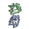
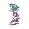
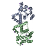
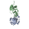
Entry Database : PDB / ID : 4b93Title Complex of Vamp7 cytoplasmic domain with 2nd ankyrin repeat domain of Varp ANKYRIN REPEAT DOMAIN-CONTAINING PROTEIN 27 VESICLE-ASSOCIATED MEMBRANE PROTEIN 7 Keywords / / Function / homology Function Domain/homology Component
/ / / / / / / / / / / / / / / / / / / / / / / / / / / / / / / / / / / / / / / / / / / / / / / / / / / / / / / / / / / / / / / / / / / / / / / / / / / / / / / / / / / / / / / / / / / / / / / / / / / / / / / / / / / / / / / / / / Biological species MUS MUSCULUS (house mouse)HOMO SAPIENS (human)Method / / / Resolution : 2 Å Authors Schaefer, I.B. / Owen, D.J. / Luzio, J.P. / Evans, P.R. Journal : Nat.Struct.Mol.Biol. / Year : 2012Title : The Binding of Varp to Vamp7 Traps Vamp7 in a Closed, Fusogenically Inactive Conformation.Authors : Schafer, I.B. / Hesketh, G.G. / Bright, N.A. / Gray, S.R. / Pryor, P.R. / Evans, P.R. / Luzio, J.P. / Owen, D.J. History Deposition Aug 31, 2012 Deposition site / Processing site Revision 1.0 Oct 31, 2012 Provider / Type Revision 1.1 Dec 19, 2012 Group Revision 1.2 Jan 25, 2017 Group Revision 1.3 Apr 3, 2019 Group / Experimental preparation / OtherCategory / pdbx_database_proc / pdbx_database_statusItem / _pdbx_database_status.recvd_author_approvalRevision 1.4 May 8, 2019 Group / Experimental preparation / Category / struct_biol / Item Revision 1.5 May 8, 2024 Group / Database references / OtherCategory chem_comp_atom / chem_comp_bond ... chem_comp_atom / chem_comp_bond / database_2 / pdbx_database_status Item / _database_2.pdbx_database_accession / _pdbx_database_status.status_code_sf
Show all Show less
 Movie
Movie Controller

 Yorodumi
 Open data
 Basic information
 Components
 Keywords
 Function and homology information
 HOMO SAPIENS (human)
 X-RAY DIFFRACTION /
  Authors
 Citation
 Structure visualization
 Downloads & links
 Other downloads
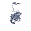


 PDBj
PDBj


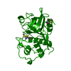
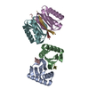
 Assembly

 Mass: 18.015 Da / Num. of mol.: 209 / Source method: isolated from a natural source / Formula: H2O
 Sample preparation
 / Beamline: ID23-1 / Wavelength: 1
  Processing