Type: MAR CCD 165 mm / Detector: CCD / Date: Sep 30, 2011
Radiation
Protocol: SINGLE WAVELENGTH / Monochromatic (M) / Laue (L): M / Scattering type: x-ray
Radiation wavelength
Wavelength: 0.9763 Å / Relative weight: 1
Reflection
Number: 179776 / Rmerge(I) obs: 0.031 / D res high: 1.4 Å / Num. obs: 67717 / % possible obs: 81.3
Diffraction reflection shell
Highest resolution: 1.4 Å / Lowest resolution: 1.48 Å / Num. obs: 5298 / % possible obs: 39.8 % / Rmerge(I) obs: 1.343
Reflection
Highest resolution: 1.4 Å / Num. obs: 67717 / % possible obs: 81.3 % / Observed criterion σ(I): -3 / Biso Wilson estimate: 29.839 Å2 / Rmerge(I) obs: 0.031 / Net I/σ(I): 16.82
Reflection shell
Rmerge(I) obs: 0.013 / Diffraction-ID: 1
Resolution (Å)
Highest resolution (Å)
Mean I/σ(I) obs
Num. measured obs
Num. unique obs
% possible all
1.4-1.48
0.51
9105
5298
39.8
1.48-1.59
1.33
15960
7493
59.9
1.59-1.71
3.09
27158
10522
90
1.71-1.88
7.45
30933
10710
99.4
1.88-2.1
16.72
28056
9643
98.7
2.1-2.42
27.51
24900
8546
98.3
2.42-2.96
35.82
20856
7256
98.3
2.96-4.19
42.78
15510
5573
96.3
4.19
44.51
7298
2676
79.5
-
Phasing
Phasing
Method: molecular replacement
Phasing MR
Model details: Phaser MODE: MR_AUTO
Highest resolution
Lowest resolution
Rotation
2.5 Å
47.67 Å
Translation
2.5 Å
47.67 Å
-
Processing
Software
Name
Version
Classification
NB
XSCALE
datascaling
PHASER
2.3.0
phasing
REFMAC
5.6.0117
refinement
PDB_EXTRACT
3.14
dataextraction
ADSC
Quantum
datacollection
XDS
datareduction
XDS
datascaling
Refinement
Method to determine structure: MOLECULAR REPLACEMENT / Resolution: 1.5→42 Å / Cor.coef. Fo:Fc: 0.968 / Cor.coef. Fo:Fc free: 0.953 / WRfactor Rfree: 0.2282 / WRfactor Rwork: 0.1872 / FOM work R set: 0.8448 / SU B: 1.446 / SU ML: 0.053 / SU R Cruickshank DPI: 0.0738 / SU Rfree: 0.0787 / Cross valid method: THROUGHOUT / σ(F): 0 / ESU R: 0.074 / ESU R Free: 0.079 / Stereochemistry target values: MAXIMUM LIKELIHOOD Details: HYDROGENS HAVE BEEN USED IF PRESENT IN THE INPUT U VALUES : REFINED INDIVIDUALLY
Rfactor
Num. reflection
% reflection
Selection details
Rfree
0.218
3115
5.1 %
RANDOM
Rwork
0.1819
-
-
-
obs
0.1838
61342
90.35 %
-
Solvent computation
Ion probe radii: 0.8 Å / Shrinkage radii: 0.8 Å / VDW probe radii: 1.2 Å / Solvent model: MASK
In the structure databanks used in Yorodumi, some data are registered as the other names, "COVID-19 virus" and "2019-nCoV". Here are the details of the virus and the list of structure data.
Jan 31, 2019. EMDB accession codes are about to change! (news from PDBe EMDB page)
EMDB accession codes are about to change! (news from PDBe EMDB page)
The allocation of 4 digits for EMDB accession codes will soon come to an end. Whilst these codes will remain in use, new EMDB accession codes will include an additional digit and will expand incrementally as the available range of codes is exhausted. The current 4-digit format prefixed with “EMD-” (i.e. EMD-XXXX) will advance to a 5-digit format (i.e. EMD-XXXXX), and so on. It is currently estimated that the 4-digit codes will be depleted around Spring 2019, at which point the 5-digit format will come into force.
The EM Navigator/Yorodumi systems omit the EMD- prefix.
Related info.:Q: What is EMD? / ID/Accession-code notation in Yorodumi/EM Navigator
Yorodumi is a browser for structure data from EMDB, PDB, SASBDB, etc.
This page is also the successor to EM Navigator detail page, and also detail information page/front-end page for Omokage search.
The word "yorodu" (or yorozu) is an old Japanese word meaning "ten thousand". "mi" (miru) is to see.
Related info.:EMDB / PDB / SASBDB / Comparison of 3 databanks / Yorodumi Search / Aug 31, 2016. New EM Navigator & Yorodumi / Yorodumi Papers / Jmol/JSmol / Function and homology information / Changes in new EM Navigator and Yorodumi
 Movie
Movie Controller

 Yorodumi
 Open data
 Basic information
 Components
 Keywords
 Function and homology information
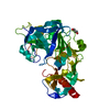
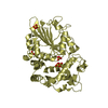
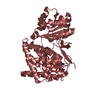
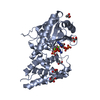
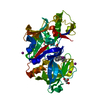
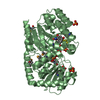
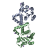
 Mycobacterium tuberculosis (bacteria)
 X-RAY DIFFRACTION /
  Authors
 Citation
 Structure visualization
 Downloads & links
 Other downloads


 PDBj
PDBj


 Assembly

 Mass: 22.990 Da / Num. of mol.: 7 / Source method: obtained synthetically / Formula: Na
 Mass: 118.174 Da / Num. of mol.: 1 / Source method: obtained synthetically / Formula: C6H14O2 / Comment: precipitant*YM
 Mass: 18.015 Da / Num. of mol.: 301 / Source method: isolated from a natural source / Formula: H2O
 Sample preparation
 / Beamline: ID23-1 / Wavelength: 0.9763 Å
 Processing