 Movie
Movie Controller

[English] 日本語
 Yorodumi
- PDB-2cu2: Crystal structure of mannose-1-phosphate geranyltransferase from ... -
+ Open data

- Basic information
| Entry | Database: PDB / ID: 2cu2 | ||||||
|---|---|---|---|---|---|---|---|
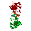
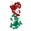
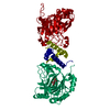
| Title | Crystal structure of mannose-1-phosphate geranyltransferase from Thermus thermophilus HB8 | ||||||
 Components Components | putative mannose-1-phosphate guanylyl transferase | ||||||
 Keywords Keywords | TRANSFERASE / mannose-1-phosphate geranyltransferase / Thermus thermophilus HB8 / Structural Genomics / RIKEN Structural Genomics/Proteomics Initiative / RSGI / NPPSFA / National Project on Protein Structural and Functional Analyses | ||||||
| Function / homology |  Function and homology information Function and homology informationmannose-1-phosphate guanylyltransferase (GTP) activity / GDP-mannose biosynthetic process / isomerase activity Similarity search - Function | ||||||
| Biological species |   Thermus thermophilus (bacteria) Thermus thermophilus (bacteria) | ||||||
| Method |  X-RAY DIFFRACTION / SYNCHROTRON / SAD / Resolution: 2.2 Å X-RAY DIFFRACTION / SYNCHROTRON / SAD / Resolution: 2.2 Å | ||||||
 Authors Authors | Sugahara, M. / Kunishima, N. / RIKEN Structural Genomics/Proteomics Initiative (RSGI) | ||||||
 Citation Citation | Journal: To be Published Title: Crystal structure of mannose-1-phosphate geranyltransferase from Thermus thermophilus HB8 Authors: Sugahara, M. / Kunishima, N. | ||||||
| History |
|
- Structure visualization
| Structure viewer | Molecule: MolmilJmol/JSmol |
|---|
- Downloads & links
-Download
| PDBx/mmCIF format | 2cu2.cif.gz | 82.1 KB | Display | PDBx/mmCIF format |
|---|---|---|---|---|
| PDB format | pdb2cu2.ent.gz | 61.6 KB | Display | PDB format |
| PDBx/mmJSON format | 2cu2.json.gz | Tree view | PDBx/mmJSON format | |
| Others |  Other downloads Other downloads |
-Validation report
| Arichive directory | https://data.pdbj.org/pub/pdb/validation_reports/cu/2cu2ftp://data.pdbj.org/pub/pdb/validation_reports/cu/2cu2 | HTTPS FTP |
|---|
-Related structure data
| Similar structure data | |
|---|---|
| Other databases |


-Links
 PDBj
PDBj

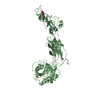
- Assembly
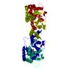
| Deposited unit | 
| |||||||||
|---|---|---|---|---|---|---|---|---|---|---|
| 1 | 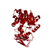
| |||||||||
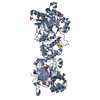
| Unit cell |
| |||||||||
| Components on special symmetry positions |
| |||||||||
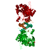
| Details | The biological assembly is a dimer in the asymmetric unit by the operations: -x+1, y, -z+1. |
-Components
| #1: Protein | Mass: 37487.867 Da / Num. of mol.: 1 Source method: isolated from a genetically manipulated source Source: (gene. exp.) Thermus thermophilus (bacteria) / Strain: HB8 / Plasmid: pET11a / Production host: References: UniProt: Q5SHI0, mannose-1-phosphate guanylyltransferase | ||
|---|---|---|---|
| #2: Chemical |   Mass: 96.063 Da / Num. of mol.: 2 / Source method: obtained synthetically / Formula: SO4 Mass: 96.063 Da / Num. of mol.: 2 / Source method: obtained synthetically / Formula: SO4#3: Water | ChemComp-HOH / |  Mass: 18.015 Da / Num. of mol.: 233 / Source method: isolated from a natural source / Formula: H2O Mass: 18.015 Da / Num. of mol.: 233 / Source method: isolated from a natural source / Formula: H2O |
-Experimental details
-Experiment
| Experiment | Method: X-RAY DIFFRACTION / Number of used crystals: 2 |
|---|
- Sample preparation
| Crystal | Density Matthews: 3.1 Å3/Da / Density % sol: 59.7 % |
|---|---|
| Crystal grow | Temperature: 295 K / Method: microbatch / pH: 8.5 Details: lithium sulphate, pH 8.5, microbatch , temperature 295K |
-Data collection
| Diffraction |
| |||||||||||||||
|---|---|---|---|---|---|---|---|---|---|---|---|---|---|---|---|---|
| Diffraction source |
| |||||||||||||||
| Detector |
| |||||||||||||||
| Radiation | Protocol: SINGLE WAVELENGTH / Monochromatic (M) / Laue (L): M / Scattering type: x-ray | |||||||||||||||
| Radiation wavelength |
| |||||||||||||||
| Reflection | Resolution: 2.2→40 Å / Num. all: 23487 / Num. obs: 23487 / % possible obs: 99.9 % / Observed criterion σ(F): 0 / Observed criterion σ(I): 0 / Redundancy: 8 % / Biso Wilson estimate: 39.749 Å2 / Rmerge(I) obs: 0.07 / Rsym value: 0.066 / Net I/σ(I): 10.1 | |||||||||||||||
| Reflection shell | Resolution: 2.2→2.28 Å / Redundancy: 8.1 % / Rmerge(I) obs: 0.62 / Mean I/σ(I) obs: 3.8 / Num. unique all: 2307 / Rsym value: 0.58 / % possible all: 100 |

- Processing
| Software |
| |||||||||||||||||||||||||
|---|---|---|---|---|---|---|---|---|---|---|---|---|---|---|---|---|---|---|---|---|---|---|---|---|---|---|
| Refinement | Method to determine structure: SAD / Resolution: 2.2→37.98 Å / Isotropic thermal model: Anisotrop / Cross valid method: THROUGHOUT / σ(F): 0 / Stereochemistry target values: Engh & Huber
| |||||||||||||||||||||||||
| Displacement parameters | Biso mean: 41.1 Å2
| |||||||||||||||||||||||||
| Refine analyze |
| |||||||||||||||||||||||||
| Refinement step | Cycle: LAST / Resolution: 2.2→37.98 Å
| |||||||||||||||||||||||||
| Refine LS restraints |
| |||||||||||||||||||||||||
| LS refinement shell | Resolution: 2.2→2.28 Å / Rfactor Rfree error: 0.028
|
