[English] 日本語
Yorodumi
- PDB-4b8l: Aurora B kinase P353G mutant -

+
Open data


ID or keywords:

Loading...

-
Basic information

Entry
Database: PDB / ID: 4b8l
TitleAurora B kinase P353G mutant
Components
  • AURORA KINASE B-A
  • INNER CENTROMERE PROTEIN A
KeywordsCELL CYCLE / CANCER
Function / homology
Function and homology information


mitotic cytokinesis checkpoint signaling / negative regulation of cytokinesis / positive regulation of mitotic sister chromatid segregation / abscission / mitotic spindle midzone assembly / cleavage furrow formation / histone H3S10 kinase activity / chromosome passenger complex / protein localization to kinetochore / negative regulation of B cell apoptotic process ...mitotic cytokinesis checkpoint signaling / negative regulation of cytokinesis / positive regulation of mitotic sister chromatid segregation / abscission / mitotic spindle midzone assembly / cleavage furrow formation / histone H3S10 kinase activity / chromosome passenger complex / protein localization to kinetochore / negative regulation of B cell apoptotic process / positive regulation of cytokinesis / mitotic cytokinesis / chromosome, centromeric region / spindle assembly / pericentric heterochromatin / post-translational protein modification / chromosome segregation / kinetochore / spindle / cellular response to UV / chromosome / midbody / microtubule / non-specific serine/threonine protein kinase / protein phosphorylation / protein serine kinase activity / protein serine/threonine kinase activity / chromatin / protein kinase binding / negative regulation of transcription by RNA polymerase II / ATP binding / metal ion binding / nucleus / cytoplasm
Similarity search - Function
Single alpha-helices involved in coiled-coils or other helix-helix interfaces - #2990 / Inner centromere protein, ARK-binding domain / Chromosome passenger complex (CPC) protein INCENP N-terminal / Inner centromere protein, ARK binding region / Chromosome passenger complex (CPC) protein INCENP N terminal / Aurora kinase / Single alpha-helices involved in coiled-coils or other helix-helix interfaces / Helix non-globular / Special / Transferase(Phosphotransferase) domain 1 ...Single alpha-helices involved in coiled-coils or other helix-helix interfaces - #2990 / Inner centromere protein, ARK-binding domain / Chromosome passenger complex (CPC) protein INCENP N-terminal / Inner centromere protein, ARK binding region / Chromosome passenger complex (CPC) protein INCENP N terminal / Aurora kinase / Single alpha-helices involved in coiled-coils or other helix-helix interfaces / Helix non-globular / Special / Transferase(Phosphotransferase) domain 1 / Transferase(Phosphotransferase); domain 1 / Phosphorylase Kinase; domain 1 / Phosphorylase Kinase; domain 1 / Serine/threonine-protein kinase, active site / Serine/Threonine protein kinases active-site signature. / Protein kinase domain / Serine/Threonine protein kinases, catalytic domain / Protein kinase, ATP binding site / Protein kinases ATP-binding region signature. / Protein kinase domain profile. / Protein kinase domain / Protein kinase-like domain superfamily / 2-Layer Sandwich / Orthogonal Bundle / Mainly Alpha / Alpha Beta
Similarity search - Domain/homology
Chem-A0P / Inner centromere protein A / Aurora kinase B-A
Similarity search - Component
Biological speciesXENOPUS LAEVIS (African clawed frog)
MethodX-RAY DIFFRACTION / SYNCHROTRON / MOLECULAR REPLACEMENT / Resolution: 3 Å
AuthorsSessa, F. / Villa, F.
Citation
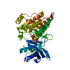
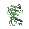
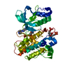
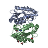
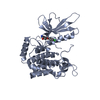
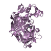
Journal: To be Published
Title: Structural and Biochemical Analysis of an Aurora B Kinase Mutant Reveals a Multistep Activation Mechanism
Authors: Sessa, F. / Villa, F.
History
DepositionAug 28, 2012Deposition site: PDBE / Processing site: PDBE
Revision 1.0Jul 10, 2013Provider: repository / Type: Initial release
Revision 1.1May 8, 2024Group: Data collection / Database references ...Data collection / Database references / Derived calculations / Other
Category: chem_comp_atom / chem_comp_bond ...chem_comp_atom / chem_comp_bond / database_2 / pdbx_database_status / struct_site
Item: _database_2.pdbx_DOI / _database_2.pdbx_database_accession ..._database_2.pdbx_DOI / _database_2.pdbx_database_accession / _pdbx_database_status.status_code_sf / _struct_site.pdbx_auth_asym_id / _struct_site.pdbx_auth_comp_id / _struct_site.pdbx_auth_seq_id

-
Structure visualization

Structure viewerMolecule:
MolmilJmol/JSmol

Downloads & links

-
Assembly

Deposited unit
A: AURORA KINASE B-A
D: INNER CENTROMERE PROTEIN A
hetero molecules


Theoretical massNumber of molelcules
Total (without water)39,1843
Polymers38,6782
Non-polymers5061
Water0
1


  • Idetical with deposited unit
  • defined by author&software
TypeNameSymmetry operationNumber
identity operation1_555x,y,z1
Buried area3810 Å2
ΔGint-23.4 kcal/mol
Surface area14840 Å2
MethodPISA
Unit cell
Length a, b, c (Å)60.896, 66.314, 89.562
Angle α, β, γ (deg.)90.00, 90.00, 90.00
Int Tables number19
Space group name H-MP212121

-
Components

#1: Protein AURORA KINASE B-A / AURORA/IPL1-RELATED KINASE 2-A / AIRK2-A / XAIRK2-A / SERINE/THREONINE-PROTEIN KINASE 12-A / ...AURORA/IPL1-RELATED KINASE 2-A / AIRK2-A / XAIRK2-A / SERINE/THREONINE-PROTEIN KINASE 12-A / SERINE/THREONINE-PROTEIN KINASE AURORA-B-A / XAURORA-B


Mass: 33603.992 Da / Num. of mol.: 1 / Fragment: RESIDUES 78-361 / Mutation: YES
Source method: isolated from a genetically manipulated source
Source: (gene. exp.) XENOPUS LAEVIS (African clawed frog) / Production host: ESCHERICHIA COLI (E. coli)
References: UniProt: Q6DE08, non-specific serine/threonine protein kinase
#2: Protein/peptide INNER CENTROMERE PROTEIN A / XL-INCENP / XINC / XINCENP / MITOTIC PHOSPHOPROTEIN 130


Mass: 5073.755 Da / Num. of mol.: 1 / Fragment: RESIDUES 797-840
Source method: isolated from a genetically manipulated source
Source: (gene. exp.) XENOPUS LAEVIS (African clawed frog) / Production host: ESCHERICHIA COLI (E. coli) / References: UniProt: O13024
#3: Chemical ChemComp-A0P / 9-{5-O-[(R)-hydroxy{[(R)-hydroxy(phosphonoamino)phosphoryl]oxy}phosphoryl]-alpha-L-arabinofuranosyl}-9H-purin-6-amine


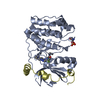
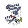
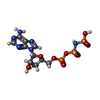
Mass: 506.196 Da / Num. of mol.: 1 / Source method: obtained synthetically / Formula: C10H17N6O12P3
Nonpolymer detailsTHE LIGAND HAS BEEN DEPOSITED UNDER THE HETEROGEN NAME A0P SEE REMARK 3 FOR DETAILS ON REFINEMENT ...THE LIGAND HAS BEEN DEPOSITED UNDER THE HETEROGEN NAME A0P SEE REMARK 3 FOR DETAILS ON REFINEMENT OF THIS COMPOUND.

-
Experimental details

-
Experiment

ExperimentMethod: X-RAY DIFFRACTION

-
Sample preparation

CrystalDensity Matthews: 2.34 Å3/Da / Density % sol: 47.39 % / Description: NONE

-
Data collection

DiffractionMean temperature: 100 K
Diffraction sourceSource: SYNCHROTRON / Site: ESRF / Beamline: ID14-3 / Wavelength: 0.934
DetectorType: ADSC CCD / Detector: CCD / Date: Jun 18, 2006
RadiationProtocol: SINGLE WAVELENGTH / Monochromatic (M) / Laue (L): M / Scattering type: x-ray
Radiation wavelengthWavelength: 0.934 Å / Relative weight: 1
ReflectionResolution: 3→25 Å / Num. obs: 7248 / % possible obs: 99 % / Observed criterion σ(I): 2 / Redundancy: 3.5 % / Rmerge(I) obs: 0.05 / Net I/σ(I): 19.5
Reflection shellResolution: 3→3.09 Å / Redundancy: 3.5 % / Rmerge(I) obs: 0.35 / Mean I/σ(I) obs: 3.8 / % possible all: 95.4

-
Processing

Software
NameVersionClassification
REFMAC5.2.0019refinement
HKL-2000data reduction
SCALEPACKdata scaling
MOLREPphasing
RefinementMethod to determine structure: MOLECULAR REPLACEMENT / Resolution: 3→50 Å / Cor.coef. Fo:Fc: 0.914 / Cor.coef. Fo:Fc free: 0.87 / SU B: 24.42 / SU ML: 0.436 / Cross valid method: THROUGHOUT / ESU R Free: 0.529 / Stereochemistry target values: MAXIMUM LIKELIHOOD
Details: HYDROGENS HAVE BEEN ADDED IN THE RIDING POSITIONS. THE AUTHORS NOTICED DURING THE REFINEMENT OF THE A0P LIGAND A DIFFERENCE IN THE CANONICAL CHIRALITY DUE TO THE TORSION OF THE LIGAND ITSELF ...Details: HYDROGENS HAVE BEEN ADDED IN THE RIDING POSITIONS. THE AUTHORS NOTICED DURING THE REFINEMENT OF THE A0P LIGAND A DIFFERENCE IN THE CANONICAL CHIRALITY DUE TO THE TORSION OF THE LIGAND ITSELF WITHIN THE ACTIVE SITE OF THE ENZYME, AND THEREFORE DECIDED TO DEPOSIT THE COORDINATES AS THEY APPEAR IN THIS ENTRY.
RfactorNum. reflection% reflectionSelection details
Rfree0.29669 347 4.6 %RANDOM
Rwork0.23684 ---
obs0.23963 7248 99.19 %-
Solvent computationIon probe radii: 0.8 Å / Shrinkage radii: 0.8 Å / VDW probe radii: 1.2 Å / Solvent model: MASK
Displacement parametersBiso mean: 65.929 Å2
Baniso -1Baniso -2Baniso -3
1-0.18 Å20 Å20 Å2
2---0.82 Å20 Å2
3---0.64 Å2
Refinement stepCycle: LAST / Resolution: 3→50 Å
ProteinNucleic acidLigandSolventTotal
Num. atoms2421 0 31 0 2452
Refine LS restraints
Refine-IDTypeDev idealDev ideal targetNumber
X-RAY DIFFRACTIONr_bond_refined_d0.0040.0222535
X-RAY DIFFRACTIONr_bond_other_d
X-RAY DIFFRACTIONr_angle_refined_deg0.8191.9833424
X-RAY DIFFRACTIONr_angle_other_deg
X-RAY DIFFRACTIONr_dihedral_angle_1_deg4.4915292
X-RAY DIFFRACTIONr_dihedral_angle_2_deg32.97923.071127
X-RAY DIFFRACTIONr_dihedral_angle_3_deg16.22115462
X-RAY DIFFRACTIONr_dihedral_angle_4_deg12.8911521
X-RAY DIFFRACTIONr_chiral_restr0.0570.2353
X-RAY DIFFRACTIONr_gen_planes_refined0.0020.021917
X-RAY DIFFRACTIONr_gen_planes_other
X-RAY DIFFRACTIONr_nbd_refined0.2310.31179
X-RAY DIFFRACTIONr_nbd_other
X-RAY DIFFRACTIONr_nbtor_refined0.330.51669
X-RAY DIFFRACTIONr_nbtor_other
X-RAY DIFFRACTIONr_xyhbond_nbd_refined0.1750.5111
X-RAY DIFFRACTIONr_xyhbond_nbd_other
X-RAY DIFFRACTIONr_metal_ion_refined
X-RAY DIFFRACTIONr_metal_ion_other
X-RAY DIFFRACTIONr_symmetry_vdw_refined0.2080.329
X-RAY DIFFRACTIONr_symmetry_vdw_other
X-RAY DIFFRACTIONr_symmetry_hbond_refined0.2290.510
X-RAY DIFFRACTIONr_symmetry_hbond_other
X-RAY DIFFRACTIONr_symmetry_metal_ion_refined
X-RAY DIFFRACTIONr_symmetry_metal_ion_other
X-RAY DIFFRACTIONr_mcbond_it0.40621483
X-RAY DIFFRACTIONr_mcbond_other
X-RAY DIFFRACTIONr_mcangle_it0.73132365
X-RAY DIFFRACTIONr_mcangle_other
X-RAY DIFFRACTIONr_scbond_it0.23121163
X-RAY DIFFRACTIONr_scbond_other
X-RAY DIFFRACTIONr_scangle_it0.41931057
X-RAY DIFFRACTIONr_scangle_other
X-RAY DIFFRACTIONr_long_range_B_refined
X-RAY DIFFRACTIONr_long_range_B_other
X-RAY DIFFRACTIONr_rigid_bond_restr
X-RAY DIFFRACTIONr_sphericity_free
X-RAY DIFFRACTIONr_sphericity_bonded
LS refinement shellResolution: 3→3.078 Å / Total num. of bins used: 20
RfactorNum. reflection% reflection
Rfree0.496 25 -
Rwork0.348 503 -
obs--97.06 %

+
About Yorodumi

-
News

-
Feb 9, 2022. New format data for meta-information of EMDB entries

New format data for meta-information of EMDB entries

  • Version 3 of the EMDB header file is now the official format.
  • The previous official version 1.9 will be removed from the archive.

Related info.:EMDB header

External links:wwPDB to switch to version 3 of the EMDB data model

-
Aug 12, 2020. Covid-19 info

Covid-19 info

URL: https://pdbj.org/emnavi/covid19.php

New page: Covid-19 featured information page in EM Navigator.

Related info.:Covid-19 info / Mar 5, 2020. Novel coronavirus structure data

+
Mar 5, 2020. Novel coronavirus structure data

Novel coronavirus structure data

Related info.:Yorodumi Speices / Aug 12, 2020. Covid-19 info

External links:COVID-19 featured content - PDBj / Molecule of the Month (242):Coronavirus Proteases

+
Jan 31, 2019. EMDB accession codes are about to change! (news from PDBe EMDB page)

EMDB accession codes are about to change! (news from PDBe EMDB page)

  • The allocation of 4 digits for EMDB accession codes will soon come to an end. Whilst these codes will remain in use, new EMDB accession codes will include an additional digit and will expand incrementally as the available range of codes is exhausted. The current 4-digit format prefixed with “EMD-” (i.e. EMD-XXXX) will advance to a 5-digit format (i.e. EMD-XXXXX), and so on. It is currently estimated that the 4-digit codes will be depleted around Spring 2019, at which point the 5-digit format will come into force.
  • The EM Navigator/Yorodumi systems omit the EMD- prefix.

Related info.:Q: What is EMD? / ID/Accession-code notation in Yorodumi/EM Navigator

External links:EMDB Accession Codes are Changing Soon! / Contact to PDBj

+
Jul 12, 2017. Major update of PDB

Major update of PDB

  • wwPDB released updated PDB data conforming to the new PDBx/mmCIF dictionary.
  • This is a major update changing the version number from 4 to 5, and with Remediation, in which all the entries are updated.
  • In this update, many items about electron microscopy experimental information are reorganized (e.g. em_software).
  • Now, EM Navigator and Yorodumi are based on the updated data.

External links:wwPDB Remediation / Enriched Model Files Conforming to OneDep Data Standards Now Available in the PDB FTP Archive

-
Yorodumi

Thousand views of thousand structures

  • Yorodumi is a browser for structure data from EMDB, PDB, SASBDB, etc.
  • This page is also the successor to EM Navigator detail page, and also detail information page/front-end page for Omokage search.
  • The word "yorodu" (or yorozu) is an old Japanese word meaning "ten thousand". "mi" (miru) is to see.

Related info.:EMDB / PDB / SASBDB / Comparison of 3 databanks / Yorodumi Search / Aug 31, 2016. New EM Navigator & Yorodumi / Yorodumi Papers / Jmol/JSmol / Function and homology information / Changes in new EM Navigator and Yorodumi

Read more