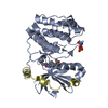
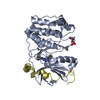
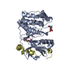
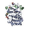
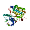
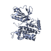
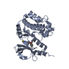
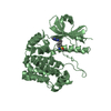
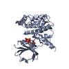
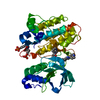
[English] 日本語
Yorodumi
- PDB-3ztx: Aurora kinase selective inhibitors identified using a Taxol-induc... -

+
Open data


ID or keywords:

Loading...

-
Basic information

Entry
Database: PDB / ID: 3ztx
TitleAurora kinase selective inhibitors identified using a Taxol-induced checkpoint sensitivity screen.
Components
  • INNER CENTROMERE PROTEIN A
  • SERINE/THREONINE-PROTEIN KINASE 12-A
KeywordsTRANSFERASE/CELL CYCLE / TRANSFERASE-CELL CYCLE COMPLEX / TRANSFERASE / TAXOL-INDUCED CHECKPOINT INHIBITOR
Function / homology
Function and homology information


mitotic cytokinesis checkpoint signaling / negative regulation of cytokinesis / positive regulation of mitotic sister chromatid segregation / abscission / mitotic spindle midzone assembly / cleavage furrow formation / histone H3S10 kinase activity / chromosome passenger complex / protein localization to kinetochore / negative regulation of B cell apoptotic process ...mitotic cytokinesis checkpoint signaling / negative regulation of cytokinesis / positive regulation of mitotic sister chromatid segregation / abscission / mitotic spindle midzone assembly / cleavage furrow formation / histone H3S10 kinase activity / chromosome passenger complex / protein localization to kinetochore / negative regulation of B cell apoptotic process / positive regulation of cytokinesis / mitotic cytokinesis / chromosome, centromeric region / spindle assembly / pericentric heterochromatin / post-translational protein modification / chromosome segregation / kinetochore / spindle / cellular response to UV / chromosome / midbody / microtubule / non-specific serine/threonine protein kinase / protein phosphorylation / protein serine kinase activity / protein serine/threonine kinase activity / chromatin / protein kinase binding / negative regulation of transcription by RNA polymerase II / ATP binding / metal ion binding / nucleus / cytoplasm
Similarity search - Function
Single alpha-helices involved in coiled-coils or other helix-helix interfaces - #2990 / Inner centromere protein, ARK-binding domain / Chromosome passenger complex (CPC) protein INCENP N-terminal / Inner centromere protein, ARK binding region / Chromosome passenger complex (CPC) protein INCENP N terminal / Aurora kinase / Single alpha-helices involved in coiled-coils or other helix-helix interfaces / Helix non-globular / Special / Transferase(Phosphotransferase) domain 1 ...Single alpha-helices involved in coiled-coils or other helix-helix interfaces - #2990 / Inner centromere protein, ARK-binding domain / Chromosome passenger complex (CPC) protein INCENP N-terminal / Inner centromere protein, ARK binding region / Chromosome passenger complex (CPC) protein INCENP N terminal / Aurora kinase / Single alpha-helices involved in coiled-coils or other helix-helix interfaces / Helix non-globular / Special / Transferase(Phosphotransferase) domain 1 / Transferase(Phosphotransferase); domain 1 / Phosphorylase Kinase; domain 1 / Phosphorylase Kinase; domain 1 / Serine/threonine-protein kinase, active site / Serine/Threonine protein kinases active-site signature. / Protein kinase domain / Serine/Threonine protein kinases, catalytic domain / Protein kinase, ATP binding site / Protein kinases ATP-binding region signature. / Protein kinase domain profile. / Protein kinase domain / Protein kinase-like domain superfamily / 2-Layer Sandwich / Orthogonal Bundle / Mainly Alpha / Alpha Beta
Similarity search - Domain/homology
Chem-ZTX / Inner centromere protein A / Aurora kinase B-A
Similarity search - Component
Biological speciesXENOPUS LAEVIS (African clawed frog)
MethodX-RAY DIFFRACTION / SYNCHROTRON / MOLECULAR REPLACEMENT / Resolution: 1.95 Å
AuthorsKwiatkowski, N. / Villa, F. / Musacchio, A. / Gray, N.
CitationJournal: Acs Chem.Biol. / Year: 2012
Title: Selective Aurora Kinase Inhibitors Identified Using a Taxol- Induced Checkpoint Sensitivity Screen.
Authors: Kwiatkowski, N. / Deng, X. / Wang, J. / Tan, L. / Villa, F. / Santaguida, S. / Huang, H.C. / Mitchison, T. / Musacchio, A. / Gray, N.
History
DepositionJul 12, 2011Deposition site: PDBE / Processing site: PDBE
Revision 1.0Feb 1, 2012Provider: repository / Type: Initial release

-
Structure visualization

Structure viewerMolecule:
MolmilJmol/JSmol

Downloads & links

-
Assembly

Deposited unit
A: SERINE/THREONINE-PROTEIN KINASE 12-A
B: SERINE/THREONINE-PROTEIN KINASE 12-A
C: INNER CENTROMERE PROTEIN A
D: INNER CENTROMERE PROTEIN A
hetero molecules


Theoretical massNumber of molelcules
Total (without water)78,0846
Polymers77,2234
Non-polymers8612
Water5,134285
1
A: SERINE/THREONINE-PROTEIN KINASE 12-A
D: INNER CENTROMERE PROTEIN A
hetero molecules


Theoretical massNumber of molelcules
Total (without water)39,0423
Polymers38,6122
Non-polymers4311
Water362
TypeNameSymmetry operationNumber
identity operation1_555x,y,z1
Buried area3330 Å2
ΔGint-24.4 kcal/mol
Surface area15570 Å2
MethodPISA
2
B: SERINE/THREONINE-PROTEIN KINASE 12-A
C: INNER CENTROMERE PROTEIN A
hetero molecules


Theoretical massNumber of molelcules
Total (without water)39,0423
Polymers38,6122
Non-polymers4311
Water362
TypeNameSymmetry operationNumber
identity operation1_555x,y,z1
Buried area3330 Å2
ΔGint-23.2 kcal/mol
Surface area15900 Å2
MethodPISA
Unit cell
Length a, b, c (Å)45.634, 66.364, 116.679
Angle α, β, γ (deg.)90.00, 96.56, 90.00
Int Tables number4
Space group name H-MP1211

-
Components

#1: Protein SERINE/THREONINE-PROTEIN KINASE 12-A / AURORA B / AURORA KINASE B-A / AURORA/IPL1-RELATED KINASE 2 / A XAIRK2 / SERINE/THREONINE-PROTEIN ...AURORA B / AURORA KINASE B-A / AURORA/IPL1-RELATED KINASE 2 / A XAIRK2 / SERINE/THREONINE-PROTEIN KINASE AURORA-B-A / XAURORA-B


Mass: 33537.871 Da / Num. of mol.: 2 / Fragment: KINASE DOMAIN, RESIDUES 78-361
Source method: isolated from a genetically manipulated source
Source: (gene. exp.) XENOPUS LAEVIS (African clawed frog) / Production host: ESCHERICHIA COLI (E. coli)
References: UniProt: Q6DE08, non-specific serine/threonine protein kinase
#2: Protein/peptide INNER CENTROMERE PROTEIN A / XL-INCENP / XINC / XINCENP / MITOTIC PHOSPHOPROTEIN 130


Mass: 5073.755 Da / Num. of mol.: 2 / Fragment: RESIDUES 797-840
Source method: isolated from a genetically manipulated source
Source: (gene. exp.) XENOPUS LAEVIS (African clawed frog) / Production host: ESCHERICHIA COLI (E. coli) / References: UniProt: O13024
#3: Chemical ChemComp-ZTX / 2-((4-(4-HYDROXYPIPERIDIN-1-YL)PHENYL)AMINO)-5,11-DIMETHYL-5H-BENZO[E]PYRIMIDO [5,4-B][1,4]DIAZEPIN-6(11H)-ONE


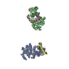
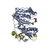
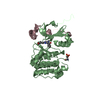
Mass: 430.502 Da / Num. of mol.: 2 / Source method: obtained synthetically / Formula: C24H26N6O2
#4: Water ChemComp-HOH / water / Water


Mass: 18.015 Da / Num. of mol.: 285 / Source method: isolated from a natural source / Formula: H2O

-
Experimental details

-
Experiment

ExperimentMethod: X-RAY DIFFRACTION / Number of used crystals: 1

-
Sample preparation

CrystalDensity Matthews: 2.28 Å3/Da / Density % sol: 46 % / Description: NONE

-
Data collection

DiffractionMean temperature: 100 K
Diffraction sourceSource: SYNCHROTRON / Site: ESRF / Beamline: ID14-1 / Wavelength: 0.9334
DetectorType: ADSC QUANTUM 210 / Detector: CCD
RadiationProtocol: SINGLE WAVELENGTH / Monochromatic (M) / Laue (L): M / Scattering type: x-ray
Radiation wavelengthWavelength: 0.9334 Å / Relative weight: 1
ReflectionResolution: 1.85→35 Å / Num. obs: 60077 / % possible obs: 99.5 % / Observed criterion σ(I): 3 / Redundancy: 2.8 % / Rmerge(I) obs: 0.06 / Net I/σ(I): 16.3
Reflection shellResolution: 1.85→1.92 Å / Redundancy: 2.8 % / Rmerge(I) obs: 0.4 / Mean I/σ(I) obs: 2.9 / % possible all: 99.2

-
Processing

Software
NameVersionClassification
REFMAC5.5.0072refinement
DENZOdata reduction
SCALEPACKdata scaling
MOLREPphasing
RefinementMethod to determine structure: MOLECULAR REPLACEMENT / Resolution: 1.95→34.68 Å / Cor.coef. Fo:Fc: 0.949 / Cor.coef. Fo:Fc free: 0.926 / SU B: 3.507 / SU ML: 0.102 / Cross valid method: THROUGHOUT / ESU R: 0.172 / ESU R Free: 0.153 / Stereochemistry target values: MAXIMUM LIKELIHOOD / Details: HYDROGENS HAVE BEEN ADDED IN THE RIDING POSITIONS.
RfactorNum. reflection% reflectionSelection details
Rfree0.22597 2561 5.1 %RANDOM
Rwork0.18569 ---
obs0.18775 47899 99.61 %-
Solvent computationIon probe radii: 0.8 Å / Shrinkage radii: 0.8 Å / VDW probe radii: 1.4 Å / Solvent model: MASK
Displacement parametersBiso mean: 27.02 Å2
Baniso -1Baniso -2Baniso -3
1--0.04 Å20 Å2-0.07 Å2
2--0.18 Å20 Å2
3----0.17 Å2
Refinement stepCycle: LAST / Resolution: 1.95→34.68 Å
ProteinNucleic acidLigandSolventTotal
Num. atoms5196 0 64 285 5545
Refine LS restraints
Refine-IDTypeDev idealDev ideal targetNumber
X-RAY DIFFRACTIONr_bond_refined_d0.0170.0225433
X-RAY DIFFRACTIONr_bond_other_d
X-RAY DIFFRACTIONr_angle_refined_deg1.641.9897342
X-RAY DIFFRACTIONr_angle_other_deg
X-RAY DIFFRACTIONr_dihedral_angle_1_deg5.9965629
X-RAY DIFFRACTIONr_dihedral_angle_2_deg34.92422.765264
X-RAY DIFFRACTIONr_dihedral_angle_3_deg16.41715980
X-RAY DIFFRACTIONr_dihedral_angle_4_deg17.3181549
X-RAY DIFFRACTIONr_chiral_restr0.110.2752
X-RAY DIFFRACTIONr_gen_planes_refined0.0090.0214166
X-RAY DIFFRACTIONr_gen_planes_other
X-RAY DIFFRACTIONr_nbd_refined
X-RAY DIFFRACTIONr_nbd_other
X-RAY DIFFRACTIONr_nbtor_refined
X-RAY DIFFRACTIONr_nbtor_other
X-RAY DIFFRACTIONr_xyhbond_nbd_refined
X-RAY DIFFRACTIONr_xyhbond_nbd_other
X-RAY DIFFRACTIONr_metal_ion_refined
X-RAY DIFFRACTIONr_metal_ion_other
X-RAY DIFFRACTIONr_symmetry_vdw_refined
X-RAY DIFFRACTIONr_symmetry_vdw_other
X-RAY DIFFRACTIONr_symmetry_hbond_refined
X-RAY DIFFRACTIONr_symmetry_hbond_other
X-RAY DIFFRACTIONr_symmetry_metal_ion_refined
X-RAY DIFFRACTIONr_symmetry_metal_ion_other
X-RAY DIFFRACTIONr_mcbond_it0.9911.53149
X-RAY DIFFRACTIONr_mcbond_other
X-RAY DIFFRACTIONr_mcangle_it1.79225104
X-RAY DIFFRACTIONr_mcangle_other
X-RAY DIFFRACTIONr_scbond_it2.78932284
X-RAY DIFFRACTIONr_scbond_other
X-RAY DIFFRACTIONr_scangle_it4.6364.52235
X-RAY DIFFRACTIONr_scangle_other
X-RAY DIFFRACTIONr_long_range_B_refined
X-RAY DIFFRACTIONr_long_range_B_other
X-RAY DIFFRACTIONr_rigid_bond_restr
X-RAY DIFFRACTIONr_sphericity_free
X-RAY DIFFRACTIONr_sphericity_bonded
LS refinement shellResolution: 1.95→2.001 Å / Total num. of bins used: 20
RfactorNum. reflection% reflection
Rfree0.27 158 -
Rwork0.222 3544 -
obs--99.22 %

+
About Yorodumi

-
News

-
Feb 9, 2022. New format data for meta-information of EMDB entries

New format data for meta-information of EMDB entries

  • Version 3 of the EMDB header file is now the official format.
  • The previous official version 1.9 will be removed from the archive.

Related info.:EMDB header

External links:wwPDB to switch to version 3 of the EMDB data model

-
Aug 12, 2020. Covid-19 info

Covid-19 info

URL: https://pdbj.org/emnavi/covid19.php

New page: Covid-19 featured information page in EM Navigator.

Related info.:Covid-19 info / Mar 5, 2020. Novel coronavirus structure data

+
Mar 5, 2020. Novel coronavirus structure data

Novel coronavirus structure data

Related info.:Yorodumi Speices / Aug 12, 2020. Covid-19 info

External links:COVID-19 featured content - PDBj / Molecule of the Month (242):Coronavirus Proteases

+
Jan 31, 2019. EMDB accession codes are about to change! (news from PDBe EMDB page)

EMDB accession codes are about to change! (news from PDBe EMDB page)

  • The allocation of 4 digits for EMDB accession codes will soon come to an end. Whilst these codes will remain in use, new EMDB accession codes will include an additional digit and will expand incrementally as the available range of codes is exhausted. The current 4-digit format prefixed with “EMD-” (i.e. EMD-XXXX) will advance to a 5-digit format (i.e. EMD-XXXXX), and so on. It is currently estimated that the 4-digit codes will be depleted around Spring 2019, at which point the 5-digit format will come into force.
  • The EM Navigator/Yorodumi systems omit the EMD- prefix.

Related info.:Q: What is EMD? / ID/Accession-code notation in Yorodumi/EM Navigator

External links:EMDB Accession Codes are Changing Soon! / Contact to PDBj

+
Jul 12, 2017. Major update of PDB

Major update of PDB

  • wwPDB released updated PDB data conforming to the new PDBx/mmCIF dictionary.
  • This is a major update changing the version number from 4 to 5, and with Remediation, in which all the entries are updated.
  • In this update, many items about electron microscopy experimental information are reorganized (e.g. em_software).
  • Now, EM Navigator and Yorodumi are based on the updated data.

External links:wwPDB Remediation / Enriched Model Files Conforming to OneDep Data Standards Now Available in the PDB FTP Archive

-
Yorodumi

Thousand views of thousand structures

  • Yorodumi is a browser for structure data from EMDB, PDB, SASBDB, etc.
  • This page is also the successor to EM Navigator detail page, and also detail information page/front-end page for Omokage search.
  • The word "yorodu" (or yorozu) is an old Japanese word meaning "ten thousand". "mi" (miru) is to see.

Related info.:EMDB / PDB / SASBDB / Comparison of 3 databanks / Yorodumi Search / Aug 31, 2016. New EM Navigator & Yorodumi / Yorodumi Papers / Jmol/JSmol / Function and homology information / Changes in new EM Navigator and Yorodumi

Read more