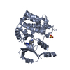
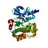
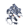
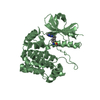
Entry Database : PDB / ID : 4zy5Title Crystal Structure of p21-activated kinase 1 in complex with an inhibitor compound 17 Serine/threonine-protein kinase PAK 1 Keywords / / / / Function / homology Function Domain/homology Component
/ / / / / / / / / / / / / / / / / / / / / / / / / / / / / / / / / / / / / / / / / / / / / / / / / / / / / / / / / / / / / / / / / / / / / / / / / / / / / / / / / / / / / / / / / / / / / / / / / / / / / / / / / / / / / / / / / / / / / / / / / / / / / / / / / Biological species Homo sapiens (human)Method / / / Resolution : 2.35 Å Authors Rouge, L. / Wang, W. Journal : J.Med.Chem. / Year : 2015Title : Structure-Guided Design of Group I Selective p21-Activated Kinase Inhibitors.Authors: Crawford, J.J. / Lee, W. / Aliagas, I. / Mathieu, S. / Hoeflich, K.P. / Zhou, W. / Wang, W. / Rouge, L. / Murray, L. / La, H. / Liu, N. / Fan, P.W. / Cheong, J. / Heise, C.E. / Ramaswamy, S. ... Authors : Crawford, J.J. / Lee, W. / Aliagas, I. / Mathieu, S. / Hoeflich, K.P. / Zhou, W. / Wang, W. / Rouge, L. / Murray, L. / La, H. / Liu, N. / Fan, P.W. / Cheong, J. / Heise, C.E. / Ramaswamy, S. / Mintzer, R. / Liu, Y. / Chao, Q. / Rudolph, J. History Deposition May 21, 2015 Deposition site / Processing site Revision 1.0 Jul 1, 2015 Provider / Type Revision 1.1 Jul 15, 2015 Group Revision 1.2 Oct 16, 2024 Group Data collection / Database references ... Data collection / Database references / Derived calculations / Source and taxonomy / Structure summary Category chem_comp_atom / chem_comp_bond ... chem_comp_atom / chem_comp_bond / citation / database_2 / entity_src_gen / pdbx_entry_details / pdbx_modification_feature / pdbx_struct_assembly / pdbx_struct_oper_list Item _citation.journal_id_CSD / _database_2.pdbx_DOI ... _citation.journal_id_CSD / _database_2.pdbx_DOI / _database_2.pdbx_database_accession / _entity_src_gen.pdbx_alt_source_flag / _pdbx_struct_assembly.oligomeric_details / _pdbx_struct_oper_list.symmetry_operation
Show all Show less
 Movie
Movie Controller

 Yorodumi
 Open data
 Basic information
 Components
 Keywords
 Function and homology information
 Homo sapiens (human)
 X-RAY DIFFRACTION /
  Authors
 Citation
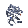
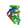
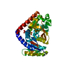
 Structure visualization
 Downloads & links
 Other downloads
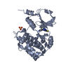


 PDBj
PDBj


 Assembly
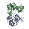


 Mass: 327.427 Da / Num. of mol.: 2 / Source method: obtained synthetically / Formula: C17H25N7
 Mass: 96.063 Da / Num. of mol.: 2 / Source method: obtained synthetically / Formula: SO4
 Mass: 78.133 Da / Num. of mol.: 1 / Source method: obtained synthetically / Formula: C2H6OS / Comment: DMSO, precipitant*YM
 Mass: 18.015 Da / Num. of mol.: 63 / Source method: isolated from a natural source / Formula: H2O
 Sample preparation
 / Beamline: 5.0.2 / Wavelength: 1 Å
 Processing