 Movie
Movie Controller

[English] 日本語
 Yorodumi
- PDB-2vgo: Crystal structure of Aurora B kinase in complex with Reversine in... -
+ Open data

- Basic information
| Entry | Database: PDB / ID: 2vgo | |||||||||
|---|---|---|---|---|---|---|---|---|---|---|
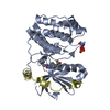
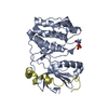
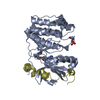
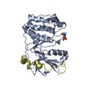
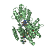
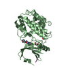
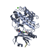
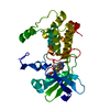
| Title | Crystal structure of Aurora B kinase in complex with Reversine inhibitor | |||||||||
 Components Components |
| |||||||||
 Keywords Keywords | TRANSFERASE / NUCLEOTIDE-BINDING / SERINE/THREONINE-PROTEIN KINASE / ATP-BINDING / COILED COIL / CELL DIVISION / KINASE / CANCER / INCENP / NUCLEUS / MITOSIS / AURORA B / METAL-BINDING / AMINOTHIAZOLE / PHOSPHORYLATION / MAGNESIUM / CELL CYCLE / CENTROMERE / MICROTUBULE | |||||||||
| Function / homology |  Function and homology information Function and homology informationmitotic cytokinesis checkpoint signaling / negative regulation of cytokinesis / positive regulation of mitotic sister chromatid segregation / meiotic spindle midzone / meiotic spindle midzone assembly / metaphase chromosome alignment / mitotic spindle midzone assembly / cleavage furrow formation / midbody abscission / protein localization to kinetochore ...mitotic cytokinesis checkpoint signaling / negative regulation of cytokinesis / positive regulation of mitotic sister chromatid segregation / meiotic spindle midzone / meiotic spindle midzone assembly / metaphase chromosome alignment / mitotic spindle midzone assembly / cleavage furrow formation / midbody abscission / protein localization to kinetochore / histone H3S10 kinase activity / chromosome passenger complex / negative regulation of B cell apoptotic process / spindle midzone / positive regulation of cytokinesis / mitotic cytokinesis / chromosome, centromeric region / pericentric heterochromatin / spindle assembly / post-translational protein modification / mitotic spindle organization / regulation of cytokinesis / spindle microtubule / chromosome segregation / kinetochore / spindle / spindle pole / cellular response to UV / chromosome / midbody / microtubule / non-specific serine/threonine protein kinase / protein serine kinase activity / protein serine/threonine kinase activity / centrosome / protein kinase binding / chromatin / negative regulation of transcription by RNA polymerase II / ATP binding / metal ion binding / nucleus Similarity search - Function | |||||||||
| Biological species | ||||||||||
| Method |  X-RAY DIFFRACTION / SYNCHROTRON / MOLECULAR REPLACEMENT / Resolution: 1.7 Å X-RAY DIFFRACTION / SYNCHROTRON / MOLECULAR REPLACEMENT / Resolution: 1.7 Å | |||||||||
 Authors Authors | D'Alise, A.M. / Amabile, G. / Iovino, M. / Di Giorgio, F.P. / Bartiromo, M. / Sessa, F. / Villa, F. / Musacchio, A. / Cortese, R. | |||||||||
 Citation Citation | Journal: Mol.Cancer Ther. / Year: 2008 Title: Reversine, a Novel Aurora Kinases Inhibitor, Inhibits Colony Formation of Human Acute Myeloid Leukemia Cells. Authors: D'Alise, A.M. / Amabile, G. / Iovino, M. / Di Giorgio, F.P. / Bartiromo, M. / Sessa, F. / Villa, F. / Musacchio, A. / Cortese, R. | |||||||||
| History |
|
- Structure visualization
| Structure viewer | Molecule: MolmilJmol/JSmol |
|---|
- Downloads & links
-Download
| PDBx/mmCIF format | 2vgo.cif.gz | 155.7 KB | Display | PDBx/mmCIF format |
|---|---|---|---|---|
| PDB format | pdb2vgo.ent.gz | 122.9 KB | Display | PDB format |
| PDBx/mmJSON format | 2vgo.json.gz | Tree view | PDBx/mmJSON format | |
| Others |  Other downloads Other downloads |
-Validation report
| Arichive directory | https://data.pdbj.org/pub/pdb/validation_reports/vg/2vgoftp://data.pdbj.org/pub/pdb/validation_reports/vg/2vgo | HTTPS FTP |
|---|
-Related structure data
| Related structure data |  2bfxS S: Starting model for refinement |
|---|---|
| Similar structure data |


-Links
 PDBj
PDBj

- Assembly
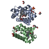
| Deposited unit | 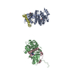
| ||||||||
|---|---|---|---|---|---|---|---|---|---|
| 1 | 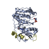
| ||||||||
| 2 | 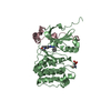
| ||||||||
| Unit cell |
|
-Components
| #1: Protein | Mass: 33537.871 Da / Num. of mol.: 2 / Fragment: RESIDUES 78-361 Source method: isolated from a genetically manipulated source Source: (gene. exp.)  References: UniProt: Q6DE08, non-specific serine/threonine protein kinase #2: Protein/peptide | Mass: 5073.755 Da / Num. of mol.: 2 / Fragment: RESIDUES 797-840 Source method: isolated from a genetically manipulated source Source: (gene. exp.) #3: Chemical |   Mass: 393.485 Da / Num. of mol.: 2 / Source method: obtained synthetically / Formula: C21H27N7O Mass: 393.485 Da / Num. of mol.: 2 / Source method: obtained synthetically / Formula: C21H27N7O#4: Water | ChemComp-HOH / |  Mass: 18.015 Da / Num. of mol.: 626 / Source method: isolated from a natural source / Formula: H2O Mass: 18.015 Da / Num. of mol.: 626 / Source method: isolated from a natural source / Formula: H2OHas protein modification | Y | |
|---|
-Experimental details
-Experiment
| Experiment | Method: X-RAY DIFFRACTION / Number of used crystals: 1 |
|---|
- Sample preparation
| Crystal | Density Matthews: 2.15 Å3/Da / Density % sol: 42.29 % / Description: NONE |
|---|
-Data collection
| Diffraction | Mean temperature: 100 K |
|---|---|
| Diffraction source | Source: SYNCHROTRON / Site: ESRF  / Beamline: ID14-1 / Wavelength: 0.9537 / Beamline: ID14-1 / Wavelength: 0.9537 |
| Detector | Type: ADSC CCD / Detector: CCD / Date: Mar 18, 2007 |
| Radiation | Protocol: SINGLE WAVELENGTH / Monochromatic (M) / Laue (L): M / Scattering type: x-ray |
| Radiation wavelength | Wavelength: 0.9537 Å / Relative weight: 1 |
| Reflection | Resolution: 1.7→116 Å / Num. obs: 72884 / % possible obs: 98.5 % / Observed criterion σ(I): 3 / Redundancy: 3.8 % / Rmerge(I) obs: 0.06 / Net I/σ(I): 20.2 |
| Reflection shell | Resolution: 1.7→1.74 Å / Redundancy: 3.6 % / Rmerge(I) obs: 0.25 / Mean I/σ(I) obs: 20.2 / % possible all: 95.5 |
- Processing
| Software |
| ||||||||||||||||||||||||||||||||||||||||||||||||||||||||||||||||||||||||||||||||||||||||||||||||||||||||||||||||||||||||||||||||||||||||||||||||||||||||||||||||||||||||||||||||||||||
|---|---|---|---|---|---|---|---|---|---|---|---|---|---|---|---|---|---|---|---|---|---|---|---|---|---|---|---|---|---|---|---|---|---|---|---|---|---|---|---|---|---|---|---|---|---|---|---|---|---|---|---|---|---|---|---|---|---|---|---|---|---|---|---|---|---|---|---|---|---|---|---|---|---|---|---|---|---|---|---|---|---|---|---|---|---|---|---|---|---|---|---|---|---|---|---|---|---|---|---|---|---|---|---|---|---|---|---|---|---|---|---|---|---|---|---|---|---|---|---|---|---|---|---|---|---|---|---|---|---|---|---|---|---|---|---|---|---|---|---|---|---|---|---|---|---|---|---|---|---|---|---|---|---|---|---|---|---|---|---|---|---|---|---|---|---|---|---|---|---|---|---|---|---|---|---|---|---|---|---|---|---|---|---|
| Refinement | Method to determine structure: MOLECULAR REPLACEMENT Starting model: PDB ENTRY 2BFX Resolution: 1.7→116.25 Å / Cor.coef. Fo:Fc: 0.951 / Cor.coef. Fo:Fc free: 0.936 / SU B: 2.132 / SU ML: 0.073 / Cross valid method: THROUGHOUT / ESU R: 0.119 / ESU R Free: 0.114 / Stereochemistry target values: MAXIMUM LIKELIHOOD / Details: HYDROGENS HAVE BEEN ADDED IN THE RIDING POSITIONS.
| ||||||||||||||||||||||||||||||||||||||||||||||||||||||||||||||||||||||||||||||||||||||||||||||||||||||||||||||||||||||||||||||||||||||||||||||||||||||||||||||||||||||||||||||||||||||
| Solvent computation | Ion probe radii: 0.8 Å / Shrinkage radii: 0.8 Å / VDW probe radii: 1.2 Å / Solvent model: MASK | ||||||||||||||||||||||||||||||||||||||||||||||||||||||||||||||||||||||||||||||||||||||||||||||||||||||||||||||||||||||||||||||||||||||||||||||||||||||||||||||||||||||||||||||||||||||
| Displacement parameters | Biso mean: 23.72 Å2
| ||||||||||||||||||||||||||||||||||||||||||||||||||||||||||||||||||||||||||||||||||||||||||||||||||||||||||||||||||||||||||||||||||||||||||||||||||||||||||||||||||||||||||||||||||||||
| Refinement step | Cycle: LAST / Resolution: 1.7→116.25 Å
| ||||||||||||||||||||||||||||||||||||||||||||||||||||||||||||||||||||||||||||||||||||||||||||||||||||||||||||||||||||||||||||||||||||||||||||||||||||||||||||||||||||||||||||||||||||||
| Refine LS restraints |
|