- PDB-4b55: Crystal Structure of the Covalent Adduct Formed between Mycobacte... -
+
Open data
ID or keywords:
Loading...
-
Basic information
Entry
Database: PDB / ID: 4b55
Title
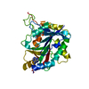
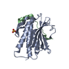
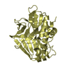
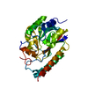
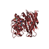
Crystal Structure of the Covalent Adduct Formed between Mycobacterium marinum Aryalamine N-acetyltransferase and Phenyl vinyl ketone a derivative of Piperidinols
Mass: 18.015 Da / Num. of mol.: 39 / Source method: isolated from a natural source / Formula: H2O
Has protein modification
Y
Nonpolymer details
3-HYDROXY-1-PHENYLPROPAN-1-ONE (RESIDUE P18) DERIVES FROM PHENYL VINYL KETONE (1-PHENYLPROP-2-EN-1- ...3-HYDROXY-1-PHENYLPROPAN-1-ONE (RESIDUE P18) DERIVES FROM PHENYL VINYL KETONE (1-PHENYLPROP-2-EN-1-ONE) (PVK). THE ADDED OH LEAVING GROUP IN THE P18 IS NOT PRESENT IN THE COMPOUND USED AS A DOUBLE BOND EXISTS BEFORE REACTION WITH CYS 70.
-
Experimental details
-
Experiment
Experiment
Method: X-RAY DIFFRACTION / Number of used crystals: 1
-
Sample preparation
Crystal
Density Matthews: 1.99 Å3/Da / Density % sol: 37 % / Description: NONE
Crystal grow
Details: 0.2 M NACL, 0.1 M NA-CACODYLATE PH 6.5 AND 2.0 M (NH4)2SO4
Resolution: 2.7→19.39 Å / Cor.coef. Fo:Fc: 0.883 / Cor.coef. Fo:Fc free: 0.7932 / Cross valid method: THROUGHOUT / σ(F): 0 / SU Rfree Blow DPI: 0.442 Details: IDEAL-DIST CONTACT TERM CONTACT SETUP. ALL ATOMS HAVE CCP4 ATOM TYPE FROM LIBRARY
Rfactor
Num. reflection
% reflection
Selection details
Rfree
0.2895
315
4.54 %
RANDOM
Rwork
0.2309
-
-
-
obs
0.2334
6944
96.78 %
-
Displacement parameters
Biso mean: 38.7 Å2
Baniso -1
Baniso -2
Baniso -3
1-
-6.8511 Å2
0 Å2
0 Å2
2-
-
-6.8511 Å2
0 Å2
3-
-
-
13.7022 Å2
Refine analyze
Luzzati coordinate error obs: 0.401 Å
Refinement step
Cycle: LAST / Resolution: 2.7→19.39 Å
Protein
Nucleic acid
Ligand
Solvent
Total
Num. atoms
2030
0
10
39
2079
Refine LS restraints
Refine-ID
Type
Dev ideal
Number
Restraint function
Weight
X-RAY DIFFRACTION
t_bond_d
0.006
2101
HARMONIC
2
X-RAY DIFFRACTION
t_angle_deg
0.82
2874
HARMONIC
2
X-RAY DIFFRACTION
t_dihedral_angle_d
681
SINUSOIDAL
2
X-RAY DIFFRACTION
t_incorr_chiral_ct
X-RAY DIFFRACTION
t_pseud_angle
X-RAY DIFFRACTION
t_trig_c_planes
40
HARMONIC
2
X-RAY DIFFRACTION
t_gen_planes
332
HARMONIC
5
X-RAY DIFFRACTION
t_it
2101
HARMONIC
20
X-RAY DIFFRACTION
t_nbd
X-RAY DIFFRACTION
t_omega_torsion
2.63
X-RAY DIFFRACTION
t_other_torsion
16.02
X-RAY DIFFRACTION
t_improper_torsion
X-RAY DIFFRACTION
t_chiral_improper_torsion
275
SEMIHARMONIC
5
X-RAY DIFFRACTION
t_sum_occupancies
X-RAY DIFFRACTION
t_utility_distance
X-RAY DIFFRACTION
t_utility_angle
X-RAY DIFFRACTION
t_utility_torsion
X-RAY DIFFRACTION
t_ideal_dist_contact
2357
SEMIHARMONIC
4
LS refinement shell
Resolution: 2.7→3.02 Å / Total num. of bins used: 5
Rfactor
Num. reflection
% reflection
Rfree
0.3098
78
4.39 %
Rwork
0.255
1699
-
all
0.2574
1777
-
obs
-
-
96.78 %
Refinement TLS params.
Method: refined / Origin x: 1.2241 Å / Origin y: 22.1945 Å / Origin z: -12.0276 Å
11
12
13
21
22
23
31
32
33
T
-0.0555 Å2
0.0113 Å2
-0.0101 Å2
-
-0.0215 Å2
0.0002 Å2
-
-
-0.1062 Å2
L
1.2961 °2
0.0908 °2
-0.0379 °2
-
0.7129 °2
-0.1555 °2
-
-
1.1535 °2
S
0.0119 Å °
0.052 Å °
0.0499 Å °
-0.0693 Å °
-0.0132 Å °
0.0491 Å °
-0.0519 Å °
-0.093 Å °
0.0012 Å °
Refinement TLS group
Selection details: CHAIN A
+
About Yorodumi
-
News
-
Feb 9, 2022. New format data for meta-information of EMDB entries
New format data for meta-information of EMDB entries
Version 3 of the EMDB header file is now the official format.
The previous official version 1.9 will be removed from the archive.
In the structure databanks used in Yorodumi, some data are registered as the other names, "COVID-19 virus" and "2019-nCoV". Here are the details of the virus and the list of structure data.
Jan 31, 2019. EMDB accession codes are about to change! (news from PDBe EMDB page)
EMDB accession codes are about to change! (news from PDBe EMDB page)
The allocation of 4 digits for EMDB accession codes will soon come to an end. Whilst these codes will remain in use, new EMDB accession codes will include an additional digit and will expand incrementally as the available range of codes is exhausted. The current 4-digit format prefixed with “EMD-” (i.e. EMD-XXXX) will advance to a 5-digit format (i.e. EMD-XXXXX), and so on. It is currently estimated that the 4-digit codes will be depleted around Spring 2019, at which point the 5-digit format will come into force.
The EM Navigator/Yorodumi systems omit the EMD- prefix.
Related info.:Q: What is EMD? / ID/Accession-code notation in Yorodumi/EM Navigator
Yorodumi is a browser for structure data from EMDB, PDB, SASBDB, etc.
This page is also the successor to EM Navigator detail page, and also detail information page/front-end page for Omokage search.
The word "yorodu" (or yorozu) is an old Japanese word meaning "ten thousand". "mi" (miru) is to see.
Related info.:EMDB / PDB / SASBDB / Comparison of 3 databanks / Yorodumi Search / Aug 31, 2016. New EM Navigator & Yorodumi / Yorodumi Papers / Jmol/JSmol / Function and homology information / Changes in new EM Navigator and Yorodumi
 Movie
Movie Controller

 Yorodumi
 Open data
 Basic information
 Components
 Keywords
 Function and homology information
 MYCOBACTERIUM MARINUM (bacteria)
 X-RAY DIFFRACTION /
  Authors
 Citation
 Structure visualization
 Downloads & links
 Other downloads
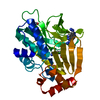


 PDBj
PDBj Assembly
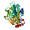
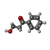
 Mass: 150.174 Da / Num. of mol.: 1 / Source method: obtained synthetically / Formula: C9H10O2
 Mass: 18.015 Da / Num. of mol.: 39 / Source method: isolated from a natural source / Formula: H2O
 Sample preparation
 / Beamline: I04 / Wavelength: 0.976
  Processing