 Movie
Movie Controller


+ Open data

- Basic information
| Entry | Database: PDB / ID: 4apc | ||||||
|---|---|---|---|---|---|---|---|
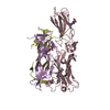
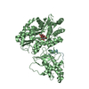
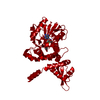
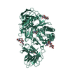
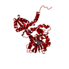
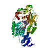
| Title | Crystal Structure of Human NIMA-related Kinase 1 (NEK1) | ||||||
 Components Components | SERINE/THREONINE-PROTEIN KINASE NEK1 | ||||||
 Keywords Keywords | TRANSFERASE | ||||||
| Function / homology |  Function and homology information Function and homology informationpericentriolar material / cilium assembly / centriolar satellite / 14-3-3 protein binding / Regulation of pyruvate metabolism / kinase activity / protein tyrosine kinase activity / protein phosphorylation / protein kinase activity / non-specific serine/threonine protein kinase ...pericentriolar material / cilium assembly / centriolar satellite / 14-3-3 protein binding / Regulation of pyruvate metabolism / kinase activity / protein tyrosine kinase activity / protein phosphorylation / protein kinase activity / non-specific serine/threonine protein kinase / protein serine kinase activity / cell division / protein serine/threonine kinase activity / centrosome / nucleoplasm / ATP binding / metal ion binding / nucleus / cytosol / cytoplasm Similarity search - Function | ||||||
| Biological species |  HOMO SAPIENS (human) HOMO SAPIENS (human) | ||||||
| Method |  X-RAY DIFFRACTION / SYNCHROTRON / MOLECULAR REPLACEMENT / Resolution: 2.1 Å X-RAY DIFFRACTION / SYNCHROTRON / MOLECULAR REPLACEMENT / Resolution: 2.1 Å | ||||||
 Authors Authors | Elkins, J.M. / Hanchuk, T.D.M. / Lovato, D.V. / Basei, F.L. / Meirelles, G.V. / Kobarg, J. / Szklarz, M. / Vollmar, M. / Mahajan, P. / Rellos, P. ...Elkins, J.M. / Hanchuk, T.D.M. / Lovato, D.V. / Basei, F.L. / Meirelles, G.V. / Kobarg, J. / Szklarz, M. / Vollmar, M. / Mahajan, P. / Rellos, P. / Zhang, Y. / Krojer, T. / Pike, A.C.W. / Bountra, C. / Arrowsmith, C. / Edwards, A. / Knapp, S. | ||||||
 Citation Citation | Journal: Sci Rep / Year: 2017 Title: NEK1 kinase domain structure and its dynamic protein interactome after exposure to Cisplatin. Authors: Melo-Hanchuk, T.D. / Slepicka, P.F. / Meirelles, G.V. / Basei, F.L. / Lovato, D.V. / Granato, D.C. / Pauletti, B.A. / Domingues, R.R. / Leme, A.F.P. / Pelegrini, A.L. / Lenz, G. / Knapp, S. ...Authors: Melo-Hanchuk, T.D. / Slepicka, P.F. / Meirelles, G.V. / Basei, F.L. / Lovato, D.V. / Granato, D.C. / Pauletti, B.A. / Domingues, R.R. / Leme, A.F.P. / Pelegrini, A.L. / Lenz, G. / Knapp, S. / Elkins, J.M. / Kobarg, J. | ||||||
| History |
|
- Structure visualization
| Structure viewer | Molecule: MolmilJmol/JSmol |
|---|
- Downloads & links
-Download
| PDBx/mmCIF format | 4apc.cif.gz | 134 KB | Display | PDBx/mmCIF format |
|---|---|---|---|---|
| PDB format | pdb4apc.ent.gz | 104.2 KB | Display | PDB format |
| PDBx/mmJSON format | 4apc.json.gz | Tree view | PDBx/mmJSON format | |
| Others |  Other downloads Other downloads |
-Validation report
| Arichive directory | https://data.pdbj.org/pub/pdb/validation_reports/ap/4apcftp://data.pdbj.org/pub/pdb/validation_reports/ap/4apc | HTTPS FTP |
|---|
-Related structure data
| Related structure data |  4b9dC  2javS S: Starting model for refinement C: citing same article ( |
|---|---|
| Similar structure data |
-Links
 PDBj
PDBj- Assembly
| Deposited unit | 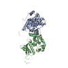
| |||||||||||||||||||||
|---|---|---|---|---|---|---|---|---|---|---|---|---|---|---|---|---|---|---|---|---|---|---|
| 1 | 
| |||||||||||||||||||||
| 2 | 
| |||||||||||||||||||||
| Unit cell |
| |||||||||||||||||||||
| Noncrystallographic symmetry (NCS) | NCS domain:
NCS domain segments: Component-ID: _ / Ens-ID: 1 / Beg auth comp-ID: HIS / Beg label comp-ID: HIS / End auth comp-ID: GLY / End label comp-ID: GLY / Refine code: _ / Auth seq-ID: -20 - 284
NCS oper: (Code: given Matrix: (0.04854, 0.99882, 0.00085), Vector: |
-Components
| #1: Protein | Mass: 39912.066 Da / Num. of mol.: 2 / Fragment: KINASE DOMAIN, RESIDUES 1-238 / Mutation: YES Source method: isolated from a genetically manipulated source Source: (gene. exp.) HOMO SAPIENS (human) / Plasmid: PNIC28-BSA4 / Production host:  References: UniProt: Q96PY6, non-specific serine/threonine protein kinase #2: Chemical | ChemComp-CL /   Mass: 35.453 Da / Num. of mol.: 7 / Source method: obtained synthetically / Formula: Cl Mass: 35.453 Da / Num. of mol.: 7 / Source method: obtained synthetically / Formula: Cl#3: Water | ChemComp-HOH / |  Mass: 18.015 Da / Num. of mol.: 165 / Source method: isolated from a natural source / Formula: H2O Mass: 18.015 Da / Num. of mol.: 165 / Source method: isolated from a natural source / Formula: H2OCompound details | ENGINEERED | Has protein modification | Y | |
|---|
-Experimental details
-Experiment
| Experiment | Method: X-RAY DIFFRACTION / Number of used crystals: 1 |
|---|
- Sample preparation
| Crystal | Density Matthews: 2.15 Å3/Da / Density % sol: 42.71 % / Description: NONE |
|---|---|
| Crystal grow | pH: 7.5 Details: 0.2 M AMMONIUM CHLORIDE, 20 %(W/V) PEG 3350, pH 7.5 |
-Data collection
| Diffraction | Mean temperature: 100 K |
|---|---|
| Diffraction source | Source: SYNCHROTRON / Site: Diamond  / Beamline: I24 / Wavelength: 0.9686 / Beamline: I24 / Wavelength: 0.9686 |
| Detector | Date: Feb 11, 2012 |
| Radiation | Protocol: SINGLE WAVELENGTH / Monochromatic (M) / Laue (L): M / Scattering type: x-ray |
| Radiation wavelength | Wavelength: 0.9686 Å / Relative weight: 1 |
| Reflection | Resolution: 2.1→46.41 Å / Num. obs: 40349 / % possible obs: 99.9 % / Observed criterion σ(I): 0 / Redundancy: 4 % / Rmerge(I) obs: 0.11 / Net I/σ(I): 8.5 |
| Reflection shell | Resolution: 2.1→2.17 Å / Redundancy: 4.1 % / Rmerge(I) obs: 0.69 / Mean I/σ(I) obs: 2.1 / % possible all: 99.9 |
- Processing
| Software |
| ||||||||||||||||||||||||||||||||||||||||||||||||||||||||||||||||||||||||||||||||||||||||||||||||||||||||||||||||||||||||||||||||||||||||||||||||||||||||||||||||||||||||||||||||||||||
|---|---|---|---|---|---|---|---|---|---|---|---|---|---|---|---|---|---|---|---|---|---|---|---|---|---|---|---|---|---|---|---|---|---|---|---|---|---|---|---|---|---|---|---|---|---|---|---|---|---|---|---|---|---|---|---|---|---|---|---|---|---|---|---|---|---|---|---|---|---|---|---|---|---|---|---|---|---|---|---|---|---|---|---|---|---|---|---|---|---|---|---|---|---|---|---|---|---|---|---|---|---|---|---|---|---|---|---|---|---|---|---|---|---|---|---|---|---|---|---|---|---|---|---|---|---|---|---|---|---|---|---|---|---|---|---|---|---|---|---|---|---|---|---|---|---|---|---|---|---|---|---|---|---|---|---|---|---|---|---|---|---|---|---|---|---|---|---|---|---|---|---|---|---|---|---|---|---|---|---|---|---|---|---|
| Refinement | Method to determine structure: MOLECULAR REPLACEMENT Starting model: PDB ENTRY 2JAV Resolution: 2.1→81.97 Å / Cor.coef. Fo:Fc: 0.934 / Cor.coef. Fo:Fc free: 0.909 / SU B: 5.061 / SU ML: 0.133 / Cross valid method: THROUGHOUT / ESU R: 0.235 / ESU R Free: 0.189 / Stereochemistry target values: MAXIMUM LIKELIHOOD / Details: HYDROGENS HAVE BEEN ADDED IN THE RIDING POSITIONS.
| ||||||||||||||||||||||||||||||||||||||||||||||||||||||||||||||||||||||||||||||||||||||||||||||||||||||||||||||||||||||||||||||||||||||||||||||||||||||||||||||||||||||||||||||||||||||
| Solvent computation | Ion probe radii: 0.8 Å / Shrinkage radii: 0.8 Å / VDW probe radii: 1.2 Å / Solvent model: MASK | ||||||||||||||||||||||||||||||||||||||||||||||||||||||||||||||||||||||||||||||||||||||||||||||||||||||||||||||||||||||||||||||||||||||||||||||||||||||||||||||||||||||||||||||||||||||
| Displacement parameters | Biso mean: 23.487 Å2
| ||||||||||||||||||||||||||||||||||||||||||||||||||||||||||||||||||||||||||||||||||||||||||||||||||||||||||||||||||||||||||||||||||||||||||||||||||||||||||||||||||||||||||||||||||||||
| Refinement step | Cycle: LAST / Resolution: 2.1→81.97 Å
| ||||||||||||||||||||||||||||||||||||||||||||||||||||||||||||||||||||||||||||||||||||||||||||||||||||||||||||||||||||||||||||||||||||||||||||||||||||||||||||||||||||||||||||||||||||||
| Refine LS restraints |
|