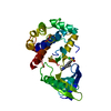
negative regulation of centriole-centriole cohesion / centrosome separation / regulation of attachment of spindle microtubules to kinetochore / regulation of mitotic centrosome separation / regulation of mitotic nuclear division / blastocyst development / positive regulation of telomere maintenance / mitotic spindle assembly / intercellular bridge / spindle assembly ...negative regulation of centriole-centriole cohesion / centrosome separation / regulation of attachment of spindle microtubules to kinetochore / regulation of mitotic centrosome separation / regulation of mitotic nuclear division / blastocyst development / positive regulation of telomere maintenance / mitotic spindle assembly / intercellular bridge / spindle assembly / Loss of Nlp from mitotic centrosomes / Loss of proteins required for interphase microtubule organization from the centrosome / Recruitment of mitotic centrosome proteins and complexes / Recruitment of NuMA to mitotic centrosomes / Anchoring of the basal body to the plasma membrane / AURKA Activation by TPX2 / APC-Cdc20 mediated degradation of Nek2A / condensed nuclear chromosome / chromosome segregation / meiotic cell cycle / kinetochore / spindle pole / Regulation of PLK1 Activity at G2/M Transition / mitotic cell cycle / midbody / protein phosphatase binding / microtubule / protein kinase activity / non-specific serine/threonine protein kinase / cilium / ciliary basal body / protein serine kinase activity / cell division / protein serine/threonine kinase activity / centrosome / nucleolus / protein-containing complex / nucleoplasm / ATP binding / metal ion binding / nucleus / plasma membrane / cytosol / cytoplasm Similarity search - Function
: / Phosphorylase Kinase; domain 1 / Phosphorylase Kinase; domain 1 / Transferase(Phosphotransferase) domain 1 / Transferase(Phosphotransferase); domain 1 / Serine/threonine-protein kinase, active site / Serine/Threonine protein kinases active-site signature. / Protein kinase domain / Serine/Threonine protein kinases, catalytic domain / Protein kinase domain profile. ...: / Phosphorylase Kinase; domain 1 / Phosphorylase Kinase; domain 1 / Transferase(Phosphotransferase) domain 1 / Transferase(Phosphotransferase); domain 1 / Serine/threonine-protein kinase, active site / Serine/Threonine protein kinases active-site signature. / Protein kinase domain / Serine/Threonine protein kinases, catalytic domain / Protein kinase domain profile. / Protein kinase domain / Protein kinase-like domain superfamily / 2-Layer Sandwich / Orthogonal Bundle / Mainly Alpha / Alpha Beta Similarity search - Domain/homology
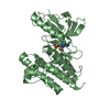
SERINE/THREONINE-PROTEINKINASENEK2 / NIMA-RELATED PROTEIN KINASE 2 / NIMA-LIKE PROTEIN KINASE 1 / HSPK 21 / NEVER IN MITOSIS GENE A- ...NIMA-RELATED PROTEIN KINASE 2 / NIMA-LIKE PROTEIN KINASE 1 / HSPK 21 / NEVER IN MITOSIS GENE A-RELATED KINASE 2
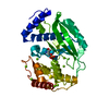
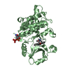
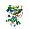
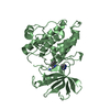
Mass: 32632.451 Da / Num. of mol.: 1 / Fragment: KINASE DOMAIN, RESIDUES 1-271 / Mutation: YES Source method: isolated from a genetically manipulated source Source: (gene. exp.) HOMO SAPIENS (human) / Plasmid: PET22B / Production host: ESCHERICHIA COLI (E. coli) / Strain (production host): BL21(DE3) / References: UniProt: P51955
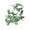
Resolution: 2.2→30 Å / Cor.coef. Fo:Fc: 0.928 / Cor.coef. Fo:Fc free: 0.883 / SU B: 11.818 / SU ML: 0.164 / TLS residual ADP flag: LIKELY RESIDUAL / Cross valid method: THROUGHOUT / ESU R: 0.286 / ESU R Free: 0.237 / Stereochemistry target values: MAXIMUM LIKELIHOOD Details: HYDROGENS HAVE BEEN ADDED IN THE RIDING POSITIONS.DIETHYLAMINO TAIL OF LIGAND IS DISORDERED AND HAS NOT BEEN INCLUDED IN THE FINAL MODEL.
Rfactor
Num. reflection
% reflection
Selection details
Rfree
0.277
796
5.1 %
RANDOM
Rwork
0.214
-
-
-
obs
0.217
14883
99.7 %
-
Solvent computation
Ion probe radii: 0.8 Å / Shrinkage radii: 0.8 Å / VDW probe radii: 1.4 Å / Solvent model: MASK
 Movie
Movie Controller


 Open data
 Basic information
 Components
 Keywords
 Function and homology information
 HOMO SAPIENS (human)
 X-RAY DIFFRACTION /
  Authors
 Citation
 Structure visualization
 Downloads & links
 Other downloads
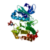


 PDBj
PDBj

 Assembly
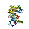


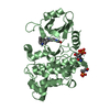
 Mass: 329.781 Da / Num. of mol.: 1 / Source method: obtained synthetically / Formula: C17H16ClN3O2
 Mass: 18.015 Da / Num. of mol.: 107 / Source method: isolated from a natural source / Formula: H2O
 Sample preparation
 / Beamline: X10SA / Wavelength: 0.95
  Processing