 Movie
Movie Controller

[English] 日本語
 Yorodumi
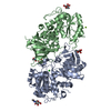
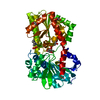
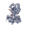
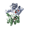
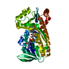
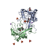
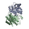
- PDB-4ap6: Crystal structure of human POFUT2 E54A mutant in complex with GDP... -
+ Open data

- Basic information
| Entry | Database: PDB / ID: 4ap6 | ||||||
|---|---|---|---|---|---|---|---|
| Title | Crystal structure of human POFUT2 E54A mutant in complex with GDP- fucose | ||||||
 Components Components | GDP-FUCOSE PROTEIN O-FUCOSYLTRANSFERASE 2 | ||||||
 Keywords Keywords | TRANSFERASE / GT-B / GT68 | ||||||
| Function / homology |  Function and homology information Function and homology informationpositive regulation of protein folding / peptide-O-fucosyltransferase / protein O-linked glycosylation via fucose / peptide-O-fucosyltransferase activity / regulation of secretion / O-glycosylation of TSR domain-containing proteins / fucose metabolic process / regulation of epithelial to mesenchymal transition / mesoderm formation / regulation of gene expression ...positive regulation of protein folding / peptide-O-fucosyltransferase / protein O-linked glycosylation via fucose / peptide-O-fucosyltransferase activity / regulation of secretion / O-glycosylation of TSR domain-containing proteins / fucose metabolic process / regulation of epithelial to mesenchymal transition / mesoderm formation / regulation of gene expression / endoplasmic reticulum membrane / Golgi apparatus Similarity search - Function | ||||||
| Biological species |  HOMO SAPIENS (human) HOMO SAPIENS (human) | ||||||
| Method |  X-RAY DIFFRACTION / SYNCHROTRON / MOLECULAR REPLACEMENT / Resolution: 3.401 Å X-RAY DIFFRACTION / SYNCHROTRON / MOLECULAR REPLACEMENT / Resolution: 3.401 Å | ||||||
 Authors Authors | Chen, C. / Keusch, J.J. / Klein, D. / Hess, D. / Hofsteenge, J. / Gut, H. | ||||||
 Citation Citation | Journal: Embo J. / Year: 2012 Title: Structure of Human Pofut2: Insights Into Thrombospondin Type 1 Repeat Fold and O-Fucosylation. Authors: Chen, C.I. / Keusch, J.J. / Klein, D. / Hess, D. / Hofsteenge, J. / Gut, H. | ||||||
| History |
|
- Structure visualization
| Structure viewer | Molecule: MolmilJmol/JSmol |
|---|
- Downloads & links
-Download
| PDBx/mmCIF format | 4ap6.cif.gz | 673.6 KB | Display | PDBx/mmCIF format |
|---|---|---|---|---|
| PDB format | pdb4ap6.ent.gz | 564 KB | Display | PDB format |
| PDBx/mmJSON format | 4ap6.json.gz | Tree view | PDBx/mmJSON format | |
| Others |  Other downloads Other downloads |
-Validation report
| Summary document | 4ap6_validation.pdf.gz | 1.5 MB | Display | wwPDB validaton report |
|---|---|---|---|---|
| Full document | 4ap6_full_validation.pdf.gz | 1.6 MB | Display | |
| Data in XML | 4ap6_validation.xml.gz | 67.3 KB | Display | |
| Data in CIF | 4ap6_validation.cif.gz | 85.2 KB | Display | |
| Arichive directory | https://data.pdbj.org/pub/pdb/validation_reports/ap/4ap6ftp://data.pdbj.org/pub/pdb/validation_reports/ap/4ap6 | HTTPS FTP |
-Related structure data


-Links
 PDBj
PDBj- Assembly
| Deposited unit | 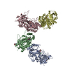
| |||||||||||||||||||||||||||||||||||||||||||||||||||||||||||||||||||||||||
|---|---|---|---|---|---|---|---|---|---|---|---|---|---|---|---|---|---|---|---|---|---|---|---|---|---|---|---|---|---|---|---|---|---|---|---|---|---|---|---|---|---|---|---|---|---|---|---|---|---|---|---|---|---|---|---|---|---|---|---|---|---|---|---|---|---|---|---|---|---|---|---|---|---|---|
| 1 | 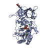
| |||||||||||||||||||||||||||||||||||||||||||||||||||||||||||||||||||||||||
| 2 | 
| |||||||||||||||||||||||||||||||||||||||||||||||||||||||||||||||||||||||||
| 3 | 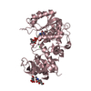
| |||||||||||||||||||||||||||||||||||||||||||||||||||||||||||||||||||||||||
| 4 | 
| |||||||||||||||||||||||||||||||||||||||||||||||||||||||||||||||||||||||||
| Unit cell |
| |||||||||||||||||||||||||||||||||||||||||||||||||||||||||||||||||||||||||
| Noncrystallographic symmetry (NCS) | NCS domain:
NCS domain segments:
NCS ensembles :
NCS oper:
|
-Components
| #1: Protein | Mass: 49553.961 Da / Num. of mol.: 4 / Fragment: RESIDUES 37-429 / Mutation: YES [E54A] Source method: isolated from a genetically manipulated source Source: (gene. exp.) HOMO SAPIENS (human) / Cell line (production host): HEK293T / Production host: HOMO SAPIENS (human) / References: UniProt: Q9Y2G5, peptide-O-fucosyltransferase#2: Chemical | ChemComp-GFB /   Mass: 589.342 Da / Num. of mol.: 4 / Source method: obtained synthetically / Formula: C16H25N5O15P2 Mass: 589.342 Da / Num. of mol.: 4 / Source method: obtained synthetically / Formula: C16H25N5O15P2#3: Sugar | ChemComp-NAG /   Type: D-saccharide, beta linking / Mass: 221.208 Da / Num. of mol.: 6 Type: D-saccharide, beta linking / Mass: 221.208 Da / Num. of mol.: 6Source method: isolated from a genetically manipulated source Formula: C8H15NO6 #4: Chemical | ChemComp-CL /   Mass: 35.453 Da / Num. of mol.: 4 / Source method: obtained synthetically / Formula: Cl Mass: 35.453 Da / Num. of mol.: 4 / Source method: obtained synthetically / Formula: Cl#5: Water | ChemComp-HOH / |  Mass: 18.015 Da / Num. of mol.: 8 / Source method: isolated from a natural source / Formula: H2O Mass: 18.015 Da / Num. of mol.: 8 / Source method: isolated from a natural source / Formula: H2OCompound details | ENGINEERED RESIDUE IN CHAIN A, GLU 54 TO ALA ENGINEERED RESIDUE IN CHAIN B, GLU 54 TO ALA ...ENGINEERED | Has protein modification | Y | |
|---|
-Experimental details
-Experiment
| Experiment | Method: X-RAY DIFFRACTION / Number of used crystals: 1 |
|---|
- Sample preparation
| Crystal | Density Matthews: 3.2 Å3/Da / Density % sol: 63 % / Description: NONE |
|---|---|
| Crystal grow | Details: 20% PEG 3350, 0.2M NASCN |
-Data collection
| Diffraction | Mean temperature: 100 K |
|---|---|
| Diffraction source | Source: SYNCHROTRON / Site: SLS  / Beamline: X06DA / Wavelength: 1 / Beamline: X06DA / Wavelength: 1 |
| Detector | Type: MARMOSAIC 225 mm CCD / Detector: CCD / Details: TOROIDAL MIRROR |
| Radiation | Monochromator: DUAL CHANNEL CUT CRYSTAL / Protocol: SINGLE WAVELENGTH / Monochromatic (M) / Laue (L): M / Scattering type: x-ray |
| Radiation wavelength | Wavelength: 1 Å / Relative weight: 1 |
| Reflection | Resolution: 3.4→40 Å / Num. obs: 32751 / % possible obs: 93.3 % / Observed criterion σ(I): -3 / Redundancy: 4.7 % / Biso Wilson estimate: 77.22 Å2 / Rmerge(I) obs: 0.17 / Net I/σ(I): 11 |
| Reflection shell | Resolution: 3.4→3.63 Å / Redundancy: 4.6 % / Rmerge(I) obs: 0.77 / Mean I/σ(I) obs: 2.1 / % possible all: 93.6 |
- Processing
| Software |
| |||||||||||||||||||||||||||||||||||||||||||||||||||||||||||||||||||||||||||||||||||||||||||||||||||||||||||||||||||||||||||||||||||||||||||||||||||||||||||||||||||||||||||||||||||||||||||||||||||||||||||||||||||||||||||||||||
|---|---|---|---|---|---|---|---|---|---|---|---|---|---|---|---|---|---|---|---|---|---|---|---|---|---|---|---|---|---|---|---|---|---|---|---|---|---|---|---|---|---|---|---|---|---|---|---|---|---|---|---|---|---|---|---|---|---|---|---|---|---|---|---|---|---|---|---|---|---|---|---|---|---|---|---|---|---|---|---|---|---|---|---|---|---|---|---|---|---|---|---|---|---|---|---|---|---|---|---|---|---|---|---|---|---|---|---|---|---|---|---|---|---|---|---|---|---|---|---|---|---|---|---|---|---|---|---|---|---|---|---|---|---|---|---|---|---|---|---|---|---|---|---|---|---|---|---|---|---|---|---|---|---|---|---|---|---|---|---|---|---|---|---|---|---|---|---|---|---|---|---|---|---|---|---|---|---|---|---|---|---|---|---|---|---|---|---|---|---|---|---|---|---|---|---|---|---|---|---|---|---|---|---|---|---|---|---|---|---|---|---|---|---|---|---|---|---|---|---|---|---|---|---|---|---|---|
| Refinement | Method to determine structure: MOLECULAR REPLACEMENT / Resolution: 3.401→39.686 Å / SU ML: 0.89 / σ(F): 1.99 / Phase error: 22 / Stereochemistry target values: ML Details: RESIDUES 298-302 ARE PARTIALLY DISORDERED. CHAIN D IS EITHER HIGHLY MOBILE OR NOT FULLY OCCUPIED IN THE CRYSTAL.
| |||||||||||||||||||||||||||||||||||||||||||||||||||||||||||||||||||||||||||||||||||||||||||||||||||||||||||||||||||||||||||||||||||||||||||||||||||||||||||||||||||||||||||||||||||||||||||||||||||||||||||||||||||||||||||||||||
| Solvent computation | Shrinkage radii: 0.83 Å / VDW probe radii: 1.1 Å / Solvent model: FLAT BULK SOLVENT MODEL / Bsol: 61.003 Å2 / ksol: 0.3 e/Å3 | |||||||||||||||||||||||||||||||||||||||||||||||||||||||||||||||||||||||||||||||||||||||||||||||||||||||||||||||||||||||||||||||||||||||||||||||||||||||||||||||||||||||||||||||||||||||||||||||||||||||||||||||||||||||||||||||||
| Displacement parameters |
| |||||||||||||||||||||||||||||||||||||||||||||||||||||||||||||||||||||||||||||||||||||||||||||||||||||||||||||||||||||||||||||||||||||||||||||||||||||||||||||||||||||||||||||||||||||||||||||||||||||||||||||||||||||||||||||||||
| Refinement step | Cycle: LAST / Resolution: 3.401→39.686 Å
| |||||||||||||||||||||||||||||||||||||||||||||||||||||||||||||||||||||||||||||||||||||||||||||||||||||||||||||||||||||||||||||||||||||||||||||||||||||||||||||||||||||||||||||||||||||||||||||||||||||||||||||||||||||||||||||||||
| Refine LS restraints |
| |||||||||||||||||||||||||||||||||||||||||||||||||||||||||||||||||||||||||||||||||||||||||||||||||||||||||||||||||||||||||||||||||||||||||||||||||||||||||||||||||||||||||||||||||||||||||||||||||||||||||||||||||||||||||||||||||
| Refine LS restraints NCS |
| |||||||||||||||||||||||||||||||||||||||||||||||||||||||||||||||||||||||||||||||||||||||||||||||||||||||||||||||||||||||||||||||||||||||||||||||||||||||||||||||||||||||||||||||||||||||||||||||||||||||||||||||||||||||||||||||||
| LS refinement shell |
| |||||||||||||||||||||||||||||||||||||||||||||||||||||||||||||||||||||||||||||||||||||||||||||||||||||||||||||||||||||||||||||||||||||||||||||||||||||||||||||||||||||||||||||||||||||||||||||||||||||||||||||||||||||||||||||||||
| Refinement TLS params. | Method: refined / Refine-ID: X-RAY DIFFRACTION
| |||||||||||||||||||||||||||||||||||||||||||||||||||||||||||||||||||||||||||||||||||||||||||||||||||||||||||||||||||||||||||||||||||||||||||||||||||||||||||||||||||||||||||||||||||||||||||||||||||||||||||||||||||||||||||||||||
| Refinement TLS group |
|
