 Movie
Movie Controller

[English] 日本語
 Yorodumi
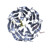
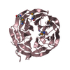
- PDB-4aow: Crystal structure of the human Rack1 protein at a resolution of 2... -
+ Open data

- Basic information
| Entry | Database: PDB / ID: 4aow | ||||||
|---|---|---|---|---|---|---|---|
| Title | Crystal structure of the human Rack1 protein at a resolution of 2.45 angstrom | ||||||
 Components Components | GUANINE NUCLEOTIDE-BINDING PROTEIN SUBUNIT BETA-2-LIKE 1 | ||||||
 Keywords Keywords | RECEPTOR / WD-REPEAT / BETA-PROPELLER | ||||||
| Function / homology |  Function and homology information Function and homology informationnegative regulation of endoplasmic reticulum unfolded protein response / positive regulation of gastrulation / protein tyrosine kinase inhibitor activity / IRE1-RACK1-PP2A complex / positive regulation of Golgi to plasma membrane protein transport / TNFR1-mediated ceramide production / cysteine-type endopeptidase activator activity involved in apoptotic process / negative regulation of intrinsic apoptotic signaling pathway in response to hydrogen peroxide / regulation of establishment of cell polarity / negative regulation of phagocytosis ...negative regulation of endoplasmic reticulum unfolded protein response / positive regulation of gastrulation / protein tyrosine kinase inhibitor activity / IRE1-RACK1-PP2A complex / positive regulation of Golgi to plasma membrane protein transport / TNFR1-mediated ceramide production / cysteine-type endopeptidase activator activity involved in apoptotic process / negative regulation of intrinsic apoptotic signaling pathway in response to hydrogen peroxide / regulation of establishment of cell polarity / negative regulation of phagocytosis / ion channel inhibitor activity / pigmentation / positive regulation of mitochondrial depolarization / negative regulation of Wnt signaling pathway / BH3 domain binding / negative regulation of translational frameshifting / regulation of adenylate cyclase-activating G protein-coupled receptor signaling pathway / positive regulation of GTPase activity / regulation of cell division / negative regulation of protein binding / protein serine/threonine kinase inhibitor activity / phagocytic cup / positive regulation of intrinsic apoptotic signaling pathway / translation regulator activity / gastrulation / signaling adaptor activity / SH2 domain binding / rescue of stalled cytosolic ribosome / cyclin binding / negative regulation of phosphatidylinositol 3-kinase/protein kinase B signal transduction / protein kinase C binding / TNFR1-induced NF-kappa-B signaling pathway / negative regulation of smoothened signaling pathway / cellular response to glucose stimulus / Regulation of TNFR1 signaling / positive regulation of protein-containing complex assembly / negative regulation of cell growth / receptor tyrosine kinase binding / positive regulation of protein phosphorylation / cellular response to growth factor stimulus / Degradation of CDH1 / enzyme activator activity / rhythmic process / regulation of protein localization / positive regulation of proteasomal ubiquitin-dependent protein catabolic process / ribosome binding / small ribosomal subunit / cytosolic small ribosomal subunit / midbody / protein phosphatase binding / molecular adaptor activity / perikaryon / cytoplasmic translation / regulation of cell cycle / negative regulation of translation / positive regulation of cell migration / protein ubiquitination / cadherin binding / positive regulation of apoptotic process / signaling receptor binding / negative regulation of gene expression / neuronal cell body / dendrite / perinuclear region of cytoplasm / enzyme binding / protein homodimerization activity / mitochondrion / RNA binding / extracellular exosome / nucleoplasm / identical protein binding / nucleus / cytoplasm / cytosol Similarity search - Function | ||||||
| Biological species |  HOMO SAPIENS (human) HOMO SAPIENS (human) | ||||||
| Method |  X-RAY DIFFRACTION / SYNCHROTRON / MOLECULAR REPLACEMENT / Resolution: 2.45 Å X-RAY DIFFRACTION / SYNCHROTRON / MOLECULAR REPLACEMENT / Resolution: 2.45 Å | ||||||
 Authors Authors | Ruiz Carrillo, D. / Chandrasekaran, R. / Nilsson, M. / Cornvick, T. / Liew, C.W. / Tan, S.M. / Lescar, J. | ||||||
 Citation Citation | Journal: Acta Crystallogr.,Sect.F / Year: 2012 Title: Structure of Human Rack1 Protein at a Resolution of 2.45 A. Authors: Ruiz Carrillo, D. / Chandrasekaran, R. / Nilsson, M. / Cornvik, T. / Liew, C.W. / Tan, S.M. / Lescar, J. | ||||||
| History |
|
- Structure visualization
| Structure viewer | Molecule: MolmilJmol/JSmol |
|---|
- Downloads & links
-Download
| PDBx/mmCIF format | 4aow.cif.gz | 360.6 KB | Display | PDBx/mmCIF format |
|---|---|---|---|---|
| PDB format | pdb4aow.ent.gz | 295.4 KB | Display | PDB format |
| PDBx/mmJSON format | 4aow.json.gz | Tree view | PDBx/mmJSON format | |
| Others |  Other downloads Other downloads |
-Validation report
| Arichive directory | https://data.pdbj.org/pub/pdb/validation_reports/ao/4aowftp://data.pdbj.org/pub/pdb/validation_reports/ao/4aow | HTTPS FTP |
|---|
-Related structure data
| Related structure data | 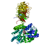 3dm0S S: Starting model for refinement |
|---|---|
| Similar structure data |


-Links
 PDBj
PDBj


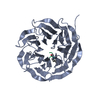
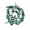
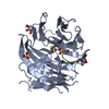
- Assembly
| Deposited unit | 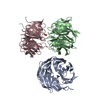
| ||||||||||||
|---|---|---|---|---|---|---|---|---|---|---|---|---|---|
| 1 | 
| ||||||||||||
| 2 | 
| ||||||||||||
| 3 | 
| ||||||||||||
| Unit cell |
| ||||||||||||
| Noncrystallographic symmetry (NCS) | NCS oper:
|
-Components
| #1: Protein | Mass: 37805.586 Da / Num. of mol.: 3 Source method: isolated from a genetically manipulated source Source: (gene. exp.) HOMO SAPIENS (human) / Plasmid: PNIC28-BSA4 / Production host:  #2: Chemical |   Mass: 92.094 Da / Num. of mol.: 2 / Source method: obtained synthetically / Formula: C3H8O3 Mass: 92.094 Da / Num. of mol.: 2 / Source method: obtained synthetically / Formula: C3H8O3#3: Water | ChemComp-HOH / |  Mass: 18.015 Da / Num. of mol.: 185 / Source method: isolated from a natural source / Formula: H2O Mass: 18.015 Da / Num. of mol.: 185 / Source method: isolated from a natural source / Formula: H2O |
|---|
-Experimental details
-Experiment
| Experiment | Method: X-RAY DIFFRACTION / Number of used crystals: 1 |
|---|
- Sample preparation
| Crystal | Density Matthews: 2.69 Å3/Da / Density % sol: 54.36 % / Description: NONE |
|---|---|
| Crystal grow | pH: 8.5 / Details: pH 8.5 |
-Data collection
| Diffraction | Mean temperature: 287 K |
|---|---|
| Diffraction source | Source: SYNCHROTRON / Site: SLS  / Beamline: X06DA / Wavelength: 1 / Beamline: X06DA / Wavelength: 1 |
| Detector | Type: MARMOSAIC 225 mm CCD / Detector: CCD / Date: Oct 23, 2011 |
| Radiation | Protocol: SINGLE WAVELENGTH / Monochromatic (M) / Laue (L): M / Scattering type: x-ray |
| Radiation wavelength | Wavelength: 1 Å / Relative weight: 1 |
| Reflection | Resolution: 2.45→47.63 Å / Num. obs: 45262 / % possible obs: 98.8 % / Observed criterion σ(I): 2.5 / Redundancy: 9.3 % / Biso Wilson estimate: 50.28 Å2 / Rmerge(I) obs: 0.11 / Net I/σ(I): 15.4 |
| Reflection shell | Resolution: 2.45→2.59 Å / Redundancy: 7.9 % / Rmerge(I) obs: 0.75 / Mean I/σ(I) obs: 2.5 / % possible all: 92.1 |
- Processing
| Software |
| |||||||||||||||||||||||||||||||||||||||||||||||||||||||||||||||||||||||||||||||||||||||||||||||||||||||||||||||||||||||||||||||||||||||||||||||||||||||||||||||||||||||||||||||
|---|---|---|---|---|---|---|---|---|---|---|---|---|---|---|---|---|---|---|---|---|---|---|---|---|---|---|---|---|---|---|---|---|---|---|---|---|---|---|---|---|---|---|---|---|---|---|---|---|---|---|---|---|---|---|---|---|---|---|---|---|---|---|---|---|---|---|---|---|---|---|---|---|---|---|---|---|---|---|---|---|---|---|---|---|---|---|---|---|---|---|---|---|---|---|---|---|---|---|---|---|---|---|---|---|---|---|---|---|---|---|---|---|---|---|---|---|---|---|---|---|---|---|---|---|---|---|---|---|---|---|---|---|---|---|---|---|---|---|---|---|---|---|---|---|---|---|---|---|---|---|---|---|---|---|---|---|---|---|---|---|---|---|---|---|---|---|---|---|---|---|---|---|---|---|---|---|
| Refinement | Method to determine structure: MOLECULAR REPLACEMENT Starting model: PDB ENTRY 3DM0 Resolution: 2.45→35.87 Å / Cor.coef. Fo:Fc: 0.9078 / Cor.coef. Fo:Fc free: 0.8901 / Cross valid method: THROUGHOUT / σ(F): 0
| |||||||||||||||||||||||||||||||||||||||||||||||||||||||||||||||||||||||||||||||||||||||||||||||||||||||||||||||||||||||||||||||||||||||||||||||||||||||||||||||||||||||||||||||
| Displacement parameters | Biso mean: 45.84 Å2
| |||||||||||||||||||||||||||||||||||||||||||||||||||||||||||||||||||||||||||||||||||||||||||||||||||||||||||||||||||||||||||||||||||||||||||||||||||||||||||||||||||||||||||||||
| Refine analyze | Luzzati coordinate error obs: 0.327 Å | |||||||||||||||||||||||||||||||||||||||||||||||||||||||||||||||||||||||||||||||||||||||||||||||||||||||||||||||||||||||||||||||||||||||||||||||||||||||||||||||||||||||||||||||
| Refinement step | Cycle: LAST / Resolution: 2.45→35.87 Å
| |||||||||||||||||||||||||||||||||||||||||||||||||||||||||||||||||||||||||||||||||||||||||||||||||||||||||||||||||||||||||||||||||||||||||||||||||||||||||||||||||||||||||||||||
| Refine LS restraints |
| |||||||||||||||||||||||||||||||||||||||||||||||||||||||||||||||||||||||||||||||||||||||||||||||||||||||||||||||||||||||||||||||||||||||||||||||||||||||||||||||||||||||||||||||
| LS refinement shell | Resolution: 2.45→2.51 Å / Total num. of bins used: 20
| |||||||||||||||||||||||||||||||||||||||||||||||||||||||||||||||||||||||||||||||||||||||||||||||||||||||||||||||||||||||||||||||||||||||||||||||||||||||||||||||||||||||||||||||
| Refinement TLS params. | Method: refined / Refine-ID: X-RAY DIFFRACTION
| |||||||||||||||||||||||||||||||||||||||||||||||||||||||||||||||||||||||||||||||||||||||||||||||||||||||||||||||||||||||||||||||||||||||||||||||||||||||||||||||||||||||||||||||
| Refinement TLS group |
|
