 Movie
Movie Controller


+ Open data

- Basic information
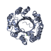
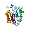
| Entry | Database: PDB / ID: 3frx | ||||||
|---|---|---|---|---|---|---|---|
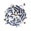
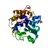
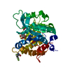
| Title | Crystal Structure of the Yeast Orthologue of RACK1, Asc1. | ||||||
 Components Components | Guanine nucleotide-binding protein subunit beta-like protein | ||||||
 Keywords Keywords | SIGNALING PROTEIN / RACK1 / WD40 / BETA PROPELLER / RIBOSOME / TRANSLATION / Acetylation / Cytoplasm / Phosphoprotein / WD repeat | ||||||
| Function / homology |  Function and homology information Function and homology informationregulation of amino acid metabolic process / negative regulation of glucose mediated signaling pathway / GDP-dissociation inhibitor activity / nonfunctional rRNA decay / ribosome-associated ubiquitin-dependent protein catabolic process / mRNA destabilization / negative regulation of translational frameshifting / G-protein alpha-subunit binding / translation regulator activity / rescue of stalled cytosolic ribosome ...regulation of amino acid metabolic process / negative regulation of glucose mediated signaling pathway / GDP-dissociation inhibitor activity / nonfunctional rRNA decay / ribosome-associated ubiquitin-dependent protein catabolic process / mRNA destabilization / negative regulation of translational frameshifting / G-protein alpha-subunit binding / translation regulator activity / rescue of stalled cytosolic ribosome / protein kinase C binding / maintenance of translational fidelity / ribosome binding / cytosolic small ribosomal subunit / cytoplasmic translation / negative regulation of translation / G protein-coupled receptor signaling pathway / negative regulation of gene expression / nucleus / cytosol / cytoplasm Similarity search - Function | ||||||
| Biological species |  | ||||||
| Method |  X-RAY DIFFRACTION / SYNCHROTRON / MOLECULAR REPLACEMENT / Resolution: 2.13 Å X-RAY DIFFRACTION / SYNCHROTRON / MOLECULAR REPLACEMENT / Resolution: 2.13 Å | ||||||
 Authors Authors | Coyle, S.M. / Gilbert, W.V. / Doudna, J.A. | ||||||
 Citation Citation | Journal: Mol.Cell.Biol. / Year: 2009 Title: Direct link between RACK1 function and localization at the ribosome in vivo Authors: Coyle, S.M. / Gilbert, W.V. / Doudna, J.A. | ||||||
| History |
|
- Structure visualization
| Structure viewer | Molecule: MolmilJmol/JSmol |
|---|
- Downloads & links
-Download
| PDBx/mmCIF format | 3frx.cif.gz | 279.1 KB | Display | PDBx/mmCIF format |
|---|---|---|---|---|
| PDB format | pdb3frx.ent.gz | 223.5 KB | Display | PDB format |
| PDBx/mmJSON format | 3frx.json.gz | Tree view | PDBx/mmJSON format | |
| Others |  Other downloads Other downloads |
-Validation report
| Arichive directory | https://data.pdbj.org/pub/pdb/validation_reports/fr/3frxftp://data.pdbj.org/pub/pdb/validation_reports/fr/3frx | HTTPS FTP |
|---|
-Related structure data
| Similar structure data |
|---|


-Links
 PDBj
PDBj

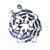
- Assembly
| Deposited unit | 
| |||||||||||||||
|---|---|---|---|---|---|---|---|---|---|---|---|---|---|---|---|---|
| 1 | 
| |||||||||||||||
| 2 | 
| |||||||||||||||
| 3 | 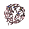
| |||||||||||||||
| 4 | 
| |||||||||||||||
| Unit cell |
| |||||||||||||||
| Components on special symmetry positions |
|
-Components
| #1: Protein | Mass: 35169.480 Da / Num. of mol.: 4 Source method: isolated from a genetically manipulated source Source: (gene. exp.) Gene: ASC1, CPC2, YM9718.15C, YMR116C / Plasmid: pSV272 / Production host:  #2: Chemical | ChemComp-MN /   Mass: 54.938 Da / Num. of mol.: 4 / Source method: obtained synthetically / Formula: Mn Mass: 54.938 Da / Num. of mol.: 4 / Source method: obtained synthetically / Formula: Mn#3: Water | ChemComp-HOH / |  Mass: 18.015 Da / Num. of mol.: 1438 / Source method: isolated from a natural source / Formula: H2O Mass: 18.015 Da / Num. of mol.: 1438 / Source method: isolated from a natural source / Formula: H2OHas protein modification | Y | |
|---|
-Experimental details
-Experiment
| Experiment | Method: X-RAY DIFFRACTION / Number of used crystals: 1 |
|---|
- Sample preparation
| Crystal | Density Matthews: 2.16 Å3/Da / Density % sol: 43.15 % |
|---|---|
| Crystal grow | Temperature: 291 K / Method: vapor diffusion, hanging drop / pH: 7.4 Details: 13.3% PEG 2000 MME, 22 mM MnOAc, 100 mM NaOAc, pH 7.4, VAPOR DIFFUSION, HANGING DROP, temperature 291K |
-Data collection
| Diffraction source | Source: SYNCHROTRON / Site: ALS  / Beamline: 8.2.1 / Wavelength: 0.9793713 Å / Beamline: 8.2.1 / Wavelength: 0.9793713 Å |
|---|---|
| Detector | Type: ADSC QUANTUM 315 / Detector: CCD / Date: Nov 16, 2007 |
| Radiation | Protocol: SINGLE WAVELENGTH / Monochromatic (M) / Laue (L): M / Scattering type: x-ray |
| Radiation wavelength | Wavelength: 0.9793713 Å / Relative weight: 1 |
| Reflection | Resolution: 2.13→17.519 Å / Num. all: 204852 / Num. obs: 125144 |
| Reflection shell | Resolution: 2.13→2.1569 Å / Num. unique all: 125144 / % possible all: 97.5 |
- Processing
| Software |
| ||||||||||||||||||||||||||||||||||||||||||||||||||||||||||||||||||||||||||||||||||||||||||||||||||||||||||||||||||||||||||||||||||||||||||||||||||||||||||||||||||||||||||||||||||||||||||||||||||||
|---|---|---|---|---|---|---|---|---|---|---|---|---|---|---|---|---|---|---|---|---|---|---|---|---|---|---|---|---|---|---|---|---|---|---|---|---|---|---|---|---|---|---|---|---|---|---|---|---|---|---|---|---|---|---|---|---|---|---|---|---|---|---|---|---|---|---|---|---|---|---|---|---|---|---|---|---|---|---|---|---|---|---|---|---|---|---|---|---|---|---|---|---|---|---|---|---|---|---|---|---|---|---|---|---|---|---|---|---|---|---|---|---|---|---|---|---|---|---|---|---|---|---|---|---|---|---|---|---|---|---|---|---|---|---|---|---|---|---|---|---|---|---|---|---|---|---|---|---|---|---|---|---|---|---|---|---|---|---|---|---|---|---|---|---|---|---|---|---|---|---|---|---|---|---|---|---|---|---|---|---|---|---|---|---|---|---|---|---|---|---|---|---|---|---|---|---|---|
| Refinement | Method to determine structure: MOLECULAR REPLACEMENT / Resolution: 2.13→17.519 Å / Occupancy max: 1 / Occupancy min: 1 / SU ML: 0.27 / σ(F): 1.2 / Phase error: 23.31 / Stereochemistry target values: ML
| ||||||||||||||||||||||||||||||||||||||||||||||||||||||||||||||||||||||||||||||||||||||||||||||||||||||||||||||||||||||||||||||||||||||||||||||||||||||||||||||||||||||||||||||||||||||||||||||||||||
| Solvent computation | Shrinkage radii: 0.9 Å / VDW probe radii: 1.11 Å / Solvent model: FLAT BULK SOLVENT MODEL / Bsol: 51.049 Å2 / ksol: 0.338 e/Å3 | ||||||||||||||||||||||||||||||||||||||||||||||||||||||||||||||||||||||||||||||||||||||||||||||||||||||||||||||||||||||||||||||||||||||||||||||||||||||||||||||||||||||||||||||||||||||||||||||||||||
| Displacement parameters | Biso max: 195.39 Å2 / Biso mean: 26.659 Å2 / Biso min: 1.47 Å2
| ||||||||||||||||||||||||||||||||||||||||||||||||||||||||||||||||||||||||||||||||||||||||||||||||||||||||||||||||||||||||||||||||||||||||||||||||||||||||||||||||||||||||||||||||||||||||||||||||||||
| Refinement step | Cycle: LAST / Resolution: 2.13→17.519 Å
| ||||||||||||||||||||||||||||||||||||||||||||||||||||||||||||||||||||||||||||||||||||||||||||||||||||||||||||||||||||||||||||||||||||||||||||||||||||||||||||||||||||||||||||||||||||||||||||||||||||
| LS refinement shell |
| ||||||||||||||||||||||||||||||||||||||||||||||||||||||||||||||||||||||||||||||||||||||||||||||||||||||||||||||||||||||||||||||||||||||||||||||||||||||||||||||||||||||||||||||||||||||||||||||||||||
| Refinement TLS params. | Method: refined / Origin x: 83.6727 Å / Origin y: 9.6898 Å / Origin z: 21.8169 Å
| ||||||||||||||||||||||||||||||||||||||||||||||||||||||||||||||||||||||||||||||||||||||||||||||||||||||||||||||||||||||||||||||||||||||||||||||||||||||||||||||||||||||||||||||||||||||||||||||||||||
| Refinement TLS group | Selection details: all |
