 Movie
Movie Controller

[English] 日本語
 Yorodumi
- PDB-4amo: Crystal Structure of the Acyltransferase Domain of the Iterative ... -
+ Open data

- Basic information
| Entry | Database: PDB / ID: 4amo | ||||||
|---|---|---|---|---|---|---|---|
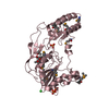
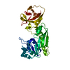
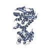
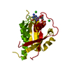
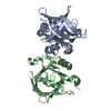
| Title | Crystal Structure of the Acyltransferase Domain of the Iterative Polyketide Synthase in Enediyne Biosynthesis Reveals the Molecular Basis of Substrate Specificity | ||||||
 Components Components | DYNE8 | ||||||
 Keywords Keywords | TRANSFERASE / ENEDIYNE / DYNEMICIN | ||||||
| Function / homology |  Function and homology information Function and homology information | ||||||
| Biological species |  MICROMONOSPORA CHERSINA (bacteria) MICROMONOSPORA CHERSINA (bacteria) | ||||||
| Method |  X-RAY DIFFRACTION / SYNCHROTRON / FOURIER SYNTHESIS / Resolution: 1.9 Å X-RAY DIFFRACTION / SYNCHROTRON / FOURIER SYNTHESIS / Resolution: 1.9 Å | ||||||
 Authors Authors | Liew, C.W. / Lescar, J. | ||||||
 Citation Citation | Journal: J.Biol.Chem. / Year: 2012 Title: Crystal Structure of the Acyltransferase Domain of the Iterative Polyketide Synthase in Enediyne Biosynthesis. Authors: Liew, C.W. / Nilsson, M. / Chen, M.W. / Sun, H. / Cornvik, T. / Liang, Z. / Lescar, J. #1: Journal: Nat.Prod.Rep. / Year: 2010 Title: Complexity and Simplicity in the Biosynthesis of Enediyne Natural Products. Authors: Liang, Z.X. | ||||||
| History |
|
- Structure visualization
| Structure viewer | Molecule: MolmilJmol/JSmol |
|---|
- Downloads & links
-Download
| PDBx/mmCIF format | 4amo.cif.gz | 86.3 KB | Display | PDBx/mmCIF format |
|---|---|---|---|---|
| PDB format | pdb4amo.ent.gz | 63.3 KB | Display | PDB format |
| PDBx/mmJSON format | 4amo.json.gz | Tree view | PDBx/mmJSON format | |
| Others |  Other downloads Other downloads |
-Validation report
| Arichive directory | https://data.pdbj.org/pub/pdb/validation_reports/am/4amoftp://data.pdbj.org/pub/pdb/validation_reports/am/4amo | HTTPS FTP |
|---|
-Related structure data
| Related structure data |  4ammSC 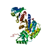 4amnC  4ampC S: Starting model for refinement C: citing same article ( |
|---|---|
| Similar structure data |


-Links
 PDBj
PDBj


- Assembly
| Deposited unit | 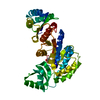
| ||||||||
|---|---|---|---|---|---|---|---|---|---|
| 1 |
| ||||||||
| Unit cell |
|
-Components
| #1: Protein | Mass: 42972.590 Da / Num. of mol.: 1 / Fragment: AT DOMAIN, RESIDUES 473 - 893 Source method: isolated from a genetically manipulated source Source: (gene. exp.) MICROMONOSPORA CHERSINA (bacteria) / Plasmid: PNIC28-BSA4-DYNE8-MICCE-EC019 / Production host: | ||||
|---|---|---|---|---|---|
| #2: Chemical |   Mass: 60.052 Da / Num. of mol.: 2 / Source method: obtained synthetically / Formula: C2H4O2 Mass: 60.052 Da / Num. of mol.: 2 / Source method: obtained synthetically / Formula: C2H4O2#3: Water | ChemComp-HOH / |  Mass: 18.015 Da / Num. of mol.: 190 / Source method: isolated from a natural source / Formula: H2O Mass: 18.015 Da / Num. of mol.: 190 / Source method: isolated from a natural source / Formula: H2OSequence details | CODON OPTIMIZED FOR E.COLI EXPRESSION | |
-Experimental details
-Experiment
| Experiment | Method: X-RAY DIFFRACTION / Number of used crystals: 5 |
|---|
- Sample preparation
| Crystal | Density Matthews: 2.07 Å3/Da / Density % sol: 40.57 % |
|---|---|
| Crystal grow | pH: 8 / Details: 0.1 M TRIS-CL PH 8.0, 30% PEG 6000 |
-Data collection
| Diffraction | Mean temperature: 287 K |
|---|---|
| Diffraction source | Source: SYNCHROTRON / Site: SLS  / Beamline: X06SA / Wavelength: 1.072 / Beamline: X06SA / Wavelength: 1.072 |
| Detector | Type: DECTRIS PILATUS 2M-F / Detector: PIXEL / Date: Apr 13, 2011 / Details: COLLIMATOR |
| Radiation | Protocol: SINGLE WAVELENGTH / Monochromatic (M) / Laue (L): M / Scattering type: x-ray |
| Radiation wavelength | Wavelength: 1.072 Å / Relative weight: 1 |
| Reflection | Resolution: 1.9→29.7 Å / Num. obs: 30505 / % possible obs: 100 % / Observed criterion σ(I): 2 / Redundancy: 10.2 % / Rmerge(I) obs: 0.08 / Net I/σ(I): 20.5 |
| Reflection shell | Resolution: 1.9→1.9 Å / Redundancy: 10.2 % / Rmerge(I) obs: 0.43 / Mean I/σ(I) obs: 5.1 / % possible all: 100 |
- Processing
| Software |
| ||||||||||||||||||||||||||||||||||||||||||||||||||||||||||||||||||||||||||||||||||||||||||||||||||||||||||||||||||||||||||||||||||||||||||||||||||||||||||||||||||||||||||||||||||||||
|---|---|---|---|---|---|---|---|---|---|---|---|---|---|---|---|---|---|---|---|---|---|---|---|---|---|---|---|---|---|---|---|---|---|---|---|---|---|---|---|---|---|---|---|---|---|---|---|---|---|---|---|---|---|---|---|---|---|---|---|---|---|---|---|---|---|---|---|---|---|---|---|---|---|---|---|---|---|---|---|---|---|---|---|---|---|---|---|---|---|---|---|---|---|---|---|---|---|---|---|---|---|---|---|---|---|---|---|---|---|---|---|---|---|---|---|---|---|---|---|---|---|---|---|---|---|---|---|---|---|---|---|---|---|---|---|---|---|---|---|---|---|---|---|---|---|---|---|---|---|---|---|---|---|---|---|---|---|---|---|---|---|---|---|---|---|---|---|---|---|---|---|---|---|---|---|---|---|---|---|---|---|---|---|
| Refinement | Method to determine structure: FOURIER SYNTHESIS Starting model: PDB ENTRY 4AMM Resolution: 1.9→29.66 Å / Cor.coef. Fo:Fc: 0.961 / Cor.coef. Fo:Fc free: 0.941 / SU B: 2.94 / SU ML: 0.089 / Cross valid method: THROUGHOUT / ESU R: 0.14 / ESU R Free: 0.133 / Stereochemistry target values: MAXIMUM LIKELIHOOD Details: HYDROGENS HAVE BEEN ADDED IN THE RIDING POSITIONS. RESIDUES FROM 586 TO 615 ARE ABSENT FROM THE MODEL OWING TO POOR ELECTRON DENSITY.
| ||||||||||||||||||||||||||||||||||||||||||||||||||||||||||||||||||||||||||||||||||||||||||||||||||||||||||||||||||||||||||||||||||||||||||||||||||||||||||||||||||||||||||||||||||||||
| Solvent computation | Ion probe radii: 0.8 Å / Shrinkage radii: 0.8 Å / VDW probe radii: 1.4 Å / Solvent model: MASK | ||||||||||||||||||||||||||||||||||||||||||||||||||||||||||||||||||||||||||||||||||||||||||||||||||||||||||||||||||||||||||||||||||||||||||||||||||||||||||||||||||||||||||||||||||||||
| Displacement parameters | Biso mean: 28.16 Å2
| ||||||||||||||||||||||||||||||||||||||||||||||||||||||||||||||||||||||||||||||||||||||||||||||||||||||||||||||||||||||||||||||||||||||||||||||||||||||||||||||||||||||||||||||||||||||
| Refinement step | Cycle: LAST / Resolution: 1.9→29.66 Å
| ||||||||||||||||||||||||||||||||||||||||||||||||||||||||||||||||||||||||||||||||||||||||||||||||||||||||||||||||||||||||||||||||||||||||||||||||||||||||||||||||||||||||||||||||||||||
| Refine LS restraints |
|