 Movie
Movie Controller

[English] 日本語
 Yorodumi
- PDB-1s3i: Crystal structure of the N terminal hydrolase domain of 10-formyl... -
+ Open data

- Basic information
| Entry | Database: PDB / ID: 1s3i | ||||||
|---|---|---|---|---|---|---|---|
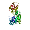
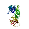
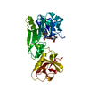
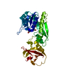
| Title | Crystal structure of the N terminal hydrolase domain of 10-formyltetrahydrofolate dehydrogenase | ||||||
 Components Components | 10-formyltetrahydrofolate dehydrogenase | ||||||
 Keywords Keywords | HYDROLASE / OXIDOREDUCTASE / Rossmann fold | ||||||
| Function / homology |  Function and homology information Function and homology informationMetabolism of folate and pterines / aldehyde dehydrogenase (NADP+) activity / formyltetrahydrofolate dehydrogenase / formyltetrahydrofolate dehydrogenase activity / 10-formyltetrahydrofolate catabolic process / aldehyde dehydrogenase [NAD(P)+] activity / NADPH regeneration / aldehyde dehydrogenase (NAD+) activity / tetrahydrofolate interconversion / folic acid metabolic process ...Metabolism of folate and pterines / aldehyde dehydrogenase (NADP+) activity / formyltetrahydrofolate dehydrogenase / formyltetrahydrofolate dehydrogenase activity / 10-formyltetrahydrofolate catabolic process / aldehyde dehydrogenase [NAD(P)+] activity / NADPH regeneration / aldehyde dehydrogenase (NAD+) activity / tetrahydrofolate interconversion / folic acid metabolic process / tetrahydrofolate biosynthetic process / protein-containing complex binding / protein-containing complex / cytosol Similarity search - Function | ||||||
| Biological species |  | ||||||
| Method |  X-RAY DIFFRACTION / MIR / Resolution: 2.3 Å X-RAY DIFFRACTION / MIR / Resolution: 2.3 Å | ||||||
 Authors Authors | Chumanevich, A.A. / Krupenko, S.A. / Davies, C. | ||||||
 Citation Citation | Journal: J.Biol.Chem. / Year: 2004 Title: The crystal structure of the hydrolase domain of 10-formyltetrahydrofolate dehydrogenase: mechanism of hydrolysis and its interplay with the dehydrogenase domain. Authors: Chumanevich, A.A. / Krupenko, S.A. / Davies, C. #1: Journal: Acta Crystallogr.,Sect.D / Year: 2002Title: Crystallization and preliminary X-ray diffraction analysis of recombinant hydrolase domain of 10-formyltetrahydrofolate dehydrogenase Authors: Chumanevich, A.A. / Davies, C. / Krupenko, S.A. | ||||||
| History |
| ||||||
| Remark 999 | SEQUENCE THE AMINO ACIDS AT POSITIONS 42, 51, 52, 283, AND 284 CONFLICT WITH THE PUBLISHED SEQUENCE. ...SEQUENCE THE AMINO ACIDS AT POSITIONS 42, 51, 52, 283, AND 284 CONFLICT WITH THE PUBLISHED SEQUENCE. THE ASPARTATE AT POSITION 310 IS NON-NATIVE AND RESULTS FROM THE CLONING. |
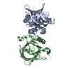
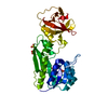
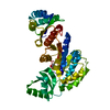
- Structure visualization
| Structure viewer | Molecule: MolmilJmol/JSmol |
|---|
- Downloads & links
-Download
| PDBx/mmCIF format | 1s3i.cif.gz | 69.1 KB | Display | PDBx/mmCIF format |
|---|---|---|---|---|
| PDB format | pdb1s3i.ent.gz | 55.5 KB | Display | PDB format |
| PDBx/mmJSON format | 1s3i.json.gz | Tree view | PDBx/mmJSON format | |
| Others |  Other downloads Other downloads |
-Validation report
| Arichive directory | https://data.pdbj.org/pub/pdb/validation_reports/s3/1s3iftp://data.pdbj.org/pub/pdb/validation_reports/s3/1s3i | HTTPS FTP |
|---|
-Related structure data
| Similar structure data |
|---|


-Links
 PDBj
PDBj


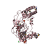
- Assembly
| Deposited unit | 
| ||||||||
|---|---|---|---|---|---|---|---|---|---|
| 1 |
| ||||||||
| Unit cell |
|
-Components
| #1: Protein | Mass: 34081.836 Da / Num. of mol.: 1 / Fragment: Nt-FDH,residues 1-310 / Mutation: none Source method: isolated from a genetically manipulated source Source: (gene. exp.)   Spodoptera frugiperda (fall armyworm) / Strain (production host): HIGH FIVE Spodoptera frugiperda (fall armyworm) / Strain (production host): HIGH FIVEReferences: UniProt: P28037, formyltetrahydrofolate dehydrogenase | ||
|---|---|---|---|
| #2: Chemical | ChemComp-BME /   Mass: 78.133 Da / Num. of mol.: 5 / Source method: obtained synthetically / Formula: C2H6OS Mass: 78.133 Da / Num. of mol.: 5 / Source method: obtained synthetically / Formula: C2H6OS#3: Water | ChemComp-HOH / |  Mass: 18.015 Da / Num. of mol.: 53 / Source method: isolated from a natural source / Formula: H2O Mass: 18.015 Da / Num. of mol.: 53 / Source method: isolated from a natural source / Formula: H2O |
-Experimental details
-Experiment
| Experiment | Method: X-RAY DIFFRACTION / Number of used crystals: 1 |
|---|
- Sample preparation
| Crystal | Density Matthews: 3.11 Å3/Da / Density % sol: 60.09 % | ||||||||||||||||||||||||
|---|---|---|---|---|---|---|---|---|---|---|---|---|---|---|---|---|---|---|---|---|---|---|---|---|---|
| Crystal grow | Temperature: 277 K / Method: vapor diffusion, sitting drop / pH: 5 Details: 1.3 M ammonium sulphate, 0.1M sodium acetate pH 5.0, VAPOR DIFFUSION, SITTING DROP, temperature 277K | ||||||||||||||||||||||||
| Crystal grow | *PLUS Temperature: 277 K / Method: vapor diffusion, sitting drop / Details: Chumanevich, A.A., (2002) Acta Cryst., D58, 1841. / PH range low: 5.1 / PH range high: 4.9 | ||||||||||||||||||||||||
| Components of the solutions | *PLUS
|
-Data collection
| Diffraction | Mean temperature: 100 K |
|---|---|
| Diffraction source | Source: ROTATING ANODE / Type: RIGAKU RU300 / Wavelength: 1.54 Å |
| Detector | Type: RIGAKU RAXIS IV / Detector: IMAGE PLATE / Date: Dec 2, 2001 / Details: mirrors |
| Radiation | Monochromator: Osmic mirrors / Protocol: SINGLE WAVELENGTH / Monochromatic (M) / Laue (L): M / Scattering type: x-ray |
| Radiation wavelength | Wavelength: 1.54 Å / Relative weight: 1 |
| Reflection | Resolution: 2.3→45.7 Å / Num. all: 19201 / Num. obs: 19201 / % possible obs: 99.7 % / Observed criterion σ(F): 0 / Observed criterion σ(I): 0 / Redundancy: 4.2 % / Biso Wilson estimate: 32.9 Å2 / Rmerge(I) obs: 0.1 / Rsym value: 0.1 / Net I/σ(I): 5.3 |
| Reflection shell | Resolution: 2.3→2.38 Å / Redundancy: 4.2 % / Rmerge(I) obs: 0.346 / Mean I/σ(I) obs: 2 / Num. unique all: 1863 / Rsym value: 0.346 / % possible all: 100 |
| Reflection | *PLUS Num. measured all: 80514 / Rmerge(I) obs: 0.1 |
| Reflection shell | *PLUS % possible obs: 100 % / Num. unique obs: 1863 / Num. measured obs: 7894 |
- Processing
| Software |
| ||||||||||||||||||||||||||||||||||||||||||||||||||||||||||||||||||||||
|---|---|---|---|---|---|---|---|---|---|---|---|---|---|---|---|---|---|---|---|---|---|---|---|---|---|---|---|---|---|---|---|---|---|---|---|---|---|---|---|---|---|---|---|---|---|---|---|---|---|---|---|---|---|---|---|---|---|---|---|---|---|---|---|---|---|---|---|---|---|---|---|
| Refinement | Method to determine structure: MIR / Resolution: 2.3→15 Å / Cor.coef. Fo:Fc: 0.915 / Cor.coef. Fo:Fc free: 0.859 / SU B: 9.584 / SU ML: 0.226 / Isotropic thermal model: isotropic / Cross valid method: THROUGHOUT / σ(F): 0 / ESU R: 0.309 / ESU R Free: 0.263 / Stereochemistry target values: MAXIMUM LIKELIHOOD
| ||||||||||||||||||||||||||||||||||||||||||||||||||||||||||||||||||||||
| Solvent computation | Ion probe radii: 0.8 Å / Shrinkage radii: 0.8 Å / VDW probe radii: 1.4 Å / Solvent model: BABINET MODEL WITH MASK | ||||||||||||||||||||||||||||||||||||||||||||||||||||||||||||||||||||||
| Displacement parameters | Biso mean: 33.745 Å2
| ||||||||||||||||||||||||||||||||||||||||||||||||||||||||||||||||||||||
| Refinement step | Cycle: LAST / Resolution: 2.3→15 Å
| ||||||||||||||||||||||||||||||||||||||||||||||||||||||||||||||||||||||
| Refine LS restraints |
| ||||||||||||||||||||||||||||||||||||||||||||||||||||||||||||||||||||||
| LS refinement shell | Resolution: 2.3→2.358 Å / Total num. of bins used: 20 /
| ||||||||||||||||||||||||||||||||||||||||||||||||||||||||||||||||||||||
| Refinement | *PLUS Highest resolution: 2.3 Å / Lowest resolution: 15 Å / Num. reflection obs: 18079 / % reflection Rfree: 5 % / Rfactor Rfree: 0.305 / Rfactor Rwork: 0.242 | ||||||||||||||||||||||||||||||||||||||||||||||||||||||||||||||||||||||
| Solvent computation | *PLUS | ||||||||||||||||||||||||||||||||||||||||||||||||||||||||||||||||||||||
| Displacement parameters | *PLUS | ||||||||||||||||||||||||||||||||||||||||||||||||||||||||||||||||||||||
| Refine LS restraints | *PLUS
| ||||||||||||||||||||||||||||||||||||||||||||||||||||||||||||||||||||||
| LS refinement shell | *PLUS Highest resolution: 2.3 Å / Lowest resolution: 2.36 Å |
