 Movie
Movie Controller

[English] 日本語
 Yorodumi
- PDB-4adu: Crystal structure of plasmodial PLP synthase with bound R5P inter... -
+ Open data

- Basic information
| Entry | Database: PDB / ID: 4adu | ||||||
|---|---|---|---|---|---|---|---|
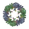
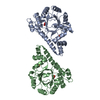
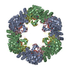
| Title | Crystal structure of plasmodial PLP synthase with bound R5P intermediate | ||||||
 Components Components | PYRIDOXINE BIOSYNTHETIC ENZYME PDX1 HOMOLOGUE, PUTATIVE | ||||||
 Keywords Keywords | TRANSFERASE / SYNTHASE / PYRIDOXAL 5-PHOSPHATE BIOSYNTHESIS | ||||||
| Function / homology | Aldolase class I / TIM Barrel / Alpha-Beta Barrel / Alpha Beta / RIBOSE-5-PHOSPHATE / :  Function and homology information Function and homology information | ||||||
| Biological species |  | ||||||
| Method |  X-RAY DIFFRACTION / SYNCHROTRON / MOLECULAR REPLACEMENT / Resolution: 2.44 Å X-RAY DIFFRACTION / SYNCHROTRON / MOLECULAR REPLACEMENT / Resolution: 2.44 Å | ||||||
 Authors Authors | Guedez, G. / Sinning, I. / Tews, I. | ||||||
 Citation Citation | Journal: Structure / Year: 2012 Title: Assembly of the Eukaryotic Plp-Synthase Complex from Plasmodium and Activation of the Pdx1 Enzyme. Authors: Guedez, G. / Hipp, K. / Windeisen, V. / Derrer, B. / Gengenbacher, M. / Boettcher, B. / Sinning, I. / Kappes, B. / Tews, I. | ||||||
| History |
| ||||||
| Remark 700 | SHEET DETERMINATION METHOD: DSSP THE SHEETS PRESENTED AS "AA" IN EACH CHAIN ON SHEET RECORDS BELOW ... SHEET DETERMINATION METHOD: DSSP THE SHEETS PRESENTED AS "AA" IN EACH CHAIN ON SHEET RECORDS BELOW IS ACTUALLY AN 8-STRANDED BARREL THIS IS REPRESENTED BY A 9-STRANDED SHEET IN WHICH THE FIRST AND LAST STRANDS ARE IDENTICAL. |
- Structure visualization
| Structure viewer | Molecule: MolmilJmol/JSmol |
|---|
- Downloads & links
-Download
| PDBx/mmCIF format | 4adu.cif.gz | 119.4 KB | Display | PDBx/mmCIF format |
|---|---|---|---|---|
| PDB format | pdb4adu.ent.gz | 93.9 KB | Display | PDB format |
| PDBx/mmJSON format | 4adu.json.gz | Tree view | PDBx/mmJSON format | |
| Others |  Other downloads Other downloads |
-Validation report
| Arichive directory | https://data.pdbj.org/pub/pdb/validation_reports/ad/4aduftp://data.pdbj.org/pub/pdb/validation_reports/ad/4adu | HTTPS FTP |
|---|
-Related structure data
| Related structure data | 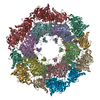 4adsC  4adtC 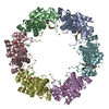 2nv1S C: citing same article ( S: Starting model for refinement |
|---|---|
| Similar structure data |


-Links
 PDBj
PDBj- Assembly
| Deposited unit | 
| ||||||||
|---|---|---|---|---|---|---|---|---|---|
| 1 | x 6
| ||||||||
| Unit cell |
|
-Components
| #1: Protein | Mass: 32733.008 Da / Num. of mol.: 2 Source method: isolated from a genetically manipulated source Source: (gene. exp.)  #2: Chemical | ChemComp-EDO /   Mass: 62.068 Da / Num. of mol.: 5 / Source method: obtained synthetically / Formula: C2H6O2 Mass: 62.068 Da / Num. of mol.: 5 / Source method: obtained synthetically / Formula: C2H6O2#3: Sugar |   Type: saccharide / Mass: 230.110 Da / Num. of mol.: 2 Type: saccharide / Mass: 230.110 Da / Num. of mol.: 2Source method: isolated from a genetically manipulated source Formula: C5H11O8P #4: Water | ChemComp-HOH / |  Mass: 18.015 Da / Num. of mol.: 153 / Source method: isolated from a natural source / Formula: H2O Mass: 18.015 Da / Num. of mol.: 153 / Source method: isolated from a natural source / Formula: H2OHas protein modification | Y | Nonpolymer details | RIBOSE 5-PHOSPHATE (R5P): LYS 84 FORMING SCHIFF BASE LINKAGE WITH R5P | |
|---|
-Experimental details
-Experiment
| Experiment | Method: X-RAY DIFFRACTION |
|---|
- Sample preparation
| Crystal | Density Matthews: 2.4 Å3/Da / Density % sol: 54 % / Description: NONE |
|---|---|
| Crystal grow | pH: 8.5 / Details: 0.1 M BICINE PH 8.5, 1.6 M LICL |
-Data collection
| Diffraction | Mean temperature: 100 K |
|---|---|
| Diffraction source | Source: SYNCHROTRON / Site: ESRF  / Beamline: ID14-4 / Wavelength: 0.88 / Beamline: ID14-4 / Wavelength: 0.88 |
| Detector | Type: ADSC CCD / Detector: CCD / Date: Jul 14, 2010 |
| Radiation | Protocol: SINGLE WAVELENGTH / Monochromatic (M) / Laue (L): M / Scattering type: x-ray |
| Radiation wavelength | Wavelength: 0.88 Å / Relative weight: 1 |
| Reflection | Resolution: 2.44→50 Å / Num. obs: 22508 / % possible obs: 99.8 % / Observed criterion σ(I): 0 / Redundancy: 6.1 % / Rmerge(I) obs: 0.07 / Net I/σ(I): 14.6 |
| Reflection shell | Resolution: 2.44→2.48 Å / Redundancy: 5.4 % / Rmerge(I) obs: 0.44 / Mean I/σ(I) obs: 2.3 / % possible all: 97.8 |
- Processing
| Software |
| ||||||||||||||||||||||||||||||||||||||||||||||||||||||||||||||||||||||||||||||||||||||||||||||||||||||||||||||||||||||||||||||||||||||||||||||||||||||||||||||||||||||||||||||||||||||
|---|---|---|---|---|---|---|---|---|---|---|---|---|---|---|---|---|---|---|---|---|---|---|---|---|---|---|---|---|---|---|---|---|---|---|---|---|---|---|---|---|---|---|---|---|---|---|---|---|---|---|---|---|---|---|---|---|---|---|---|---|---|---|---|---|---|---|---|---|---|---|---|---|---|---|---|---|---|---|---|---|---|---|---|---|---|---|---|---|---|---|---|---|---|---|---|---|---|---|---|---|---|---|---|---|---|---|---|---|---|---|---|---|---|---|---|---|---|---|---|---|---|---|---|---|---|---|---|---|---|---|---|---|---|---|---|---|---|---|---|---|---|---|---|---|---|---|---|---|---|---|---|---|---|---|---|---|---|---|---|---|---|---|---|---|---|---|---|---|---|---|---|---|---|---|---|---|---|---|---|---|---|---|---|
| Refinement | Method to determine structure: MOLECULAR REPLACEMENT Starting model: PDB ENTRY 2NV1 Resolution: 2.44→50 Å / Cor.coef. Fo:Fc: 0.948 / Cor.coef. Fo:Fc free: 0.909 / SU B: 18.164 / SU ML: 0.189 / Cross valid method: THROUGHOUT / ESU R: 0.459 / ESU R Free: 0.275 / Stereochemistry target values: MAXIMUM LIKELIHOOD / Details: HYDROGENS HAVE BEEN ADDED IN THE RIDING POSITIONS.
| ||||||||||||||||||||||||||||||||||||||||||||||||||||||||||||||||||||||||||||||||||||||||||||||||||||||||||||||||||||||||||||||||||||||||||||||||||||||||||||||||||||||||||||||||||||||
| Solvent computation | Ion probe radii: 0.8 Å / Shrinkage radii: 0.8 Å / VDW probe radii: 1.4 Å / Solvent model: BABINET MODEL WITH MASK | ||||||||||||||||||||||||||||||||||||||||||||||||||||||||||||||||||||||||||||||||||||||||||||||||||||||||||||||||||||||||||||||||||||||||||||||||||||||||||||||||||||||||||||||||||||||
| Displacement parameters | Biso mean: 39.485 Å2
| ||||||||||||||||||||||||||||||||||||||||||||||||||||||||||||||||||||||||||||||||||||||||||||||||||||||||||||||||||||||||||||||||||||||||||||||||||||||||||||||||||||||||||||||||||||||
| Refinement step | Cycle: LAST / Resolution: 2.44→50 Å
| ||||||||||||||||||||||||||||||||||||||||||||||||||||||||||||||||||||||||||||||||||||||||||||||||||||||||||||||||||||||||||||||||||||||||||||||||||||||||||||||||||||||||||||||||||||||
| Refine LS restraints |
|