 Movie
Movie Controller

[English] 日本語
 Yorodumi
- PDB-4acs: Crystal structure of mutant GST A2-2 with enhanced catalytic effi... -
+ Open data

- Basic information
| Entry | Database: PDB / ID: 4acs | ||||||
|---|---|---|---|---|---|---|---|
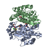
| Title | Crystal structure of mutant GST A2-2 with enhanced catalytic efficiency with azathioprine | ||||||
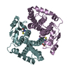
 Components Components | GLUTATHIONE S-TRANSFERASE A2 | ||||||
 Keywords Keywords | TRANSFERASE / OXIDATION-REDUCTION | ||||||
| Function / homology |  Function and homology information Function and homology informationGlutathione conjugation / Azathioprine ADME / glutathione transferase / glutathione transferase activity / epithelial cell differentiation / xenobiotic metabolic process / Gene and protein expression by JAK-STAT signaling after Interleukin-12 stimulation / glutathione metabolic process / extracellular exosome / cytosol Similarity search - Function | ||||||
| Biological species |  HOMO SAPIENS (human) HOMO SAPIENS (human) | ||||||
| Method |  X-RAY DIFFRACTION / SYNCHROTRON / MOLECULAR REPLACEMENT / Resolution: 2.1 Å X-RAY DIFFRACTION / SYNCHROTRON / MOLECULAR REPLACEMENT / Resolution: 2.1 Å | ||||||
 Authors Authors | Zhang, W. / Moden, O. / Tars, K. / Mannervik, B. | ||||||
 Citation Citation | Journal: Chem.Biol. / Year: 2012 Title: Structure-based redesign of GST A2-2 for enhanced catalytic efficiency with azathioprine. Authors: Zhang, W. / Moden, O. / Tars, K. / Mannervik, B. | ||||||
| History |
|
- Structure visualization
| Structure viewer | Molecule: MolmilJmol/JSmol |
|---|
- Downloads & links
-Download
| PDBx/mmCIF format | 4acs.cif.gz | 193.2 KB | Display | PDBx/mmCIF format |
|---|---|---|---|---|
| PDB format | pdb4acs.ent.gz | 154.9 KB | Display | PDB format |
| PDBx/mmJSON format | 4acs.json.gz | Tree view | PDBx/mmJSON format | |
| Others |  Other downloads Other downloads |
-Validation report
| Arichive directory | https://data.pdbj.org/pub/pdb/validation_reports/ac/4acsftp://data.pdbj.org/pub/pdb/validation_reports/ac/4acs | HTTPS FTP |
|---|
-Related structure data
| Related structure data |  2wjuS S: Starting model for refinement |
|---|---|
| Similar structure data |


-Links
 PDBj
PDBj


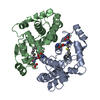
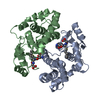
- Assembly
| Deposited unit | 
| ||||||||||||||||
|---|---|---|---|---|---|---|---|---|---|---|---|---|---|---|---|---|---|
| 1 | 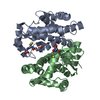
| ||||||||||||||||
| 2 | 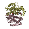
| ||||||||||||||||
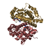
| Unit cell |
| ||||||||||||||||
| Noncrystallographic symmetry (NCS) | NCS oper:
|
-Components
| #1: Protein | Mass: 25627.738 Da / Num. of mol.: 4 / Mutation: YES Source method: isolated from a genetically manipulated source Source: (gene. exp.) HOMO SAPIENS (human) / Production host:  #2: Chemical | ChemComp-GSH /   Mass: 307.323 Da / Num. of mol.: 4 / Source method: obtained synthetically / Formula: C10H17N3O6S Mass: 307.323 Da / Num. of mol.: 4 / Source method: obtained synthetically / Formula: C10H17N3O6S#3: Water | ChemComp-HOH / |  Mass: 18.015 Da / Num. of mol.: 515 / Source method: isolated from a natural source / Formula: H2O Mass: 18.015 Da / Num. of mol.: 515 / Source method: isolated from a natural source / Formula: H2OCompound details | ENGINEERED RESIDUE IN CHAIN A, LEU 107 TO GLY ENGINEERED RESIDUE IN CHAIN A, LEU 108 TO ASP ...ENGINEERED | |
|---|
-Experimental details
-Experiment
| Experiment | Method: X-RAY DIFFRACTION / Number of used crystals: 1 |
|---|
- Sample preparation
| Crystal | Density Matthews: 2.6 Å3/Da / Density % sol: 54 % / Description: NONE |
|---|---|
| Crystal grow | Method: vapor diffusion, hanging drop / pH: 7.8 Details: CRYSTALS WERE GROWN BY HANING DROP METHOD BY MIXING EQUAL VOLUMES (4 UL) OF PROTEIN (10 MG/ML)AND GLUTATHIONE S-CONJUGATE (5 MM) MIXTURE AND RESERVOIR SOLUTION CONTAINING 9-14% PEG 4000 AND ...Details: CRYSTALS WERE GROWN BY HANING DROP METHOD BY MIXING EQUAL VOLUMES (4 UL) OF PROTEIN (10 MG/ML)AND GLUTATHIONE S-CONJUGATE (5 MM) MIXTURE AND RESERVOIR SOLUTION CONTAINING 9-14% PEG 4000 AND 2 MM DTT IN 100 MM TRIS-HCL (PH 7.8) WITH ADDITIONAL OCTYL D-BETA- GLUCOPYRANOSIDE TO A FINAL CONCENTRATION OF 0.1% (W/V) |
-Data collection
| Diffraction | Mean temperature: 100 K |
|---|---|
| Diffraction source | Source: SYNCHROTRON / Site: ESRF  / Beamline: ID14-2 / Type: ESRF / Wavelength: 0.9334 / Beamline: ID14-2 / Type: ESRF / Wavelength: 0.9334 |
| Detector | Detector: CCD / Date: Jul 14, 2010 |
| Radiation | Protocol: SINGLE WAVELENGTH / Monochromatic (M) / Laue (L): M / Scattering type: x-ray |
| Radiation wavelength | Wavelength: 0.9334 Å / Relative weight: 1 |
| Reflection | Resolution: 2.1→38 Å / Num. obs: 55710 / % possible obs: 93 % / Observed criterion σ(I): 2.6 / Redundancy: 3.2 % / Biso Wilson estimate: 13 Å2 / Rmerge(I) obs: 0.12 / Net I/σ(I): 5.1 |
| Reflection shell | Resolution: 2.1→2.2 Å / Redundancy: 2.9 % / Rmerge(I) obs: 0.29 / Mean I/σ(I) obs: 2.6 / % possible all: 77 |
- Processing
| Software |
| ||||||||||||||||||||||||||||||||||||||||||||||||||||||||||||||||||||||||||||||||||||||||||||||||||||||||||||||||||||||||||||||||||||||||||||||||||||||||||||||||||||||||||||||||||||||
|---|---|---|---|---|---|---|---|---|---|---|---|---|---|---|---|---|---|---|---|---|---|---|---|---|---|---|---|---|---|---|---|---|---|---|---|---|---|---|---|---|---|---|---|---|---|---|---|---|---|---|---|---|---|---|---|---|---|---|---|---|---|---|---|---|---|---|---|---|---|---|---|---|---|---|---|---|---|---|---|---|---|---|---|---|---|---|---|---|---|---|---|---|---|---|---|---|---|---|---|---|---|---|---|---|---|---|---|---|---|---|---|---|---|---|---|---|---|---|---|---|---|---|---|---|---|---|---|---|---|---|---|---|---|---|---|---|---|---|---|---|---|---|---|---|---|---|---|---|---|---|---|---|---|---|---|---|---|---|---|---|---|---|---|---|---|---|---|---|---|---|---|---|---|---|---|---|---|---|---|---|---|---|---|
| Refinement | Method to determine structure: MOLECULAR REPLACEMENT Starting model: PDB ENTRY 2WJU Resolution: 2.1→40 Å / Cor.coef. Fo:Fc: 0.831 / Cor.coef. Fo:Fc free: 0.756 / SU B: 7.732 / SU ML: 0.201 / Cross valid method: THROUGHOUT / ESU R: 0.071 / ESU R Free: 0.057 / Stereochemistry target values: MAXIMUM LIKELIHOOD Details: HYDROGENS HAVE BEEN ADDED IN THE RIDING POSITIONS. U VALUES REFINED INDIVIDUALLY.
| ||||||||||||||||||||||||||||||||||||||||||||||||||||||||||||||||||||||||||||||||||||||||||||||||||||||||||||||||||||||||||||||||||||||||||||||||||||||||||||||||||||||||||||||||||||||
| Solvent computation | Ion probe radii: 0.8 Å / Shrinkage radii: 0.8 Å / VDW probe radii: 1.2 Å / Solvent model: MASK | ||||||||||||||||||||||||||||||||||||||||||||||||||||||||||||||||||||||||||||||||||||||||||||||||||||||||||||||||||||||||||||||||||||||||||||||||||||||||||||||||||||||||||||||||||||||
| Displacement parameters | Biso mean: 16.155 Å2
| ||||||||||||||||||||||||||||||||||||||||||||||||||||||||||||||||||||||||||||||||||||||||||||||||||||||||||||||||||||||||||||||||||||||||||||||||||||||||||||||||||||||||||||||||||||||
| Refinement step | Cycle: LAST / Resolution: 2.1→40 Å
| ||||||||||||||||||||||||||||||||||||||||||||||||||||||||||||||||||||||||||||||||||||||||||||||||||||||||||||||||||||||||||||||||||||||||||||||||||||||||||||||||||||||||||||||||||||||
| Refine LS restraints |
|