Mass: 18.015 Da / Num. of mol.: 721 / Source method: isolated from a natural source / Formula: H2O
-
Experimental details
-
Experiment
Experiment
Method: X-RAY DIFFRACTION / Number of used crystals: 1
-
Sample preparation
Crystal
Density Matthews: 2.33 Å3/Da / Density % sol: 47.31 %
Crystal grow
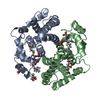
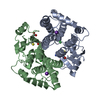
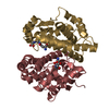
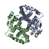
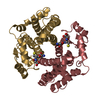
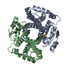
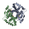
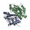
Temperature: 298 K / Method: vapor diffusion, hanging drop Details: A 4uL hanging drop comprising: 2uL 14 mg/mL R15L hGSTA1-1 solution added to 2uL reservoir buffer (5mM S-hexyl-glutathione, 0.1M TrisHCl, pH 7.5, 10 mM DTT, 15% PEG 4000) was left to ...Details: A 4uL hanging drop comprising: 2uL 14 mg/mL R15L hGSTA1-1 solution added to 2uL reservoir buffer (5mM S-hexyl-glutathione, 0.1M TrisHCl, pH 7.5, 10 mM DTT, 15% PEG 4000) was left to equilibrate in a sealed 24 plate well with 1 mL of reservoir buffer. Crystals were grown for 3 days before harvesting, VAPOR DIFFUSION, HANGING DROP, temperature 298K
-
Data collection
Diffraction
Mean temperature: 113 K
Diffraction source
Source: ROTATING ANODE / Type: RIGAKU / Wavelength: 1.5418 Å
Detector
Type: RIGAKU RAXIS IV / Detector: IMAGE PLATE / Date: Feb 13, 2006
Radiation
Monochromator: Ni / Protocol: SINGLE WAVELENGTH / Monochromatic (M) / Laue (L): M / Scattering type: x-ray
In the structure databanks used in Yorodumi, some data are registered as the other names, "COVID-19 virus" and "2019-nCoV". Here are the details of the virus and the list of structure data.
Jan 31, 2019. EMDB accession codes are about to change! (news from PDBe EMDB page)
EMDB accession codes are about to change! (news from PDBe EMDB page)
The allocation of 4 digits for EMDB accession codes will soon come to an end. Whilst these codes will remain in use, new EMDB accession codes will include an additional digit and will expand incrementally as the available range of codes is exhausted. The current 4-digit format prefixed with “EMD-” (i.e. EMD-XXXX) will advance to a 5-digit format (i.e. EMD-XXXXX), and so on. It is currently estimated that the 4-digit codes will be depleted around Spring 2019, at which point the 5-digit format will come into force.
The EM Navigator/Yorodumi systems omit the EMD- prefix.
Related info.:Q: What is EMD? / ID/Accession-code notation in Yorodumi/EM Navigator
Yorodumi is a browser for structure data from EMDB, PDB, SASBDB, etc.
This page is also the successor to EM Navigator detail page, and also detail information page/front-end page for Omokage search.
The word "yorodu" (or yorozu) is an old Japanese word meaning "ten thousand". "mi" (miru) is to see.
Related info.:EMDB / PDB / SASBDB / Comparison of 3 databanks / Yorodumi Search / Aug 31, 2016. New EM Navigator & Yorodumi / Yorodumi Papers / Jmol/JSmol / Function and homology information / Changes in new EM Navigator and Yorodumi
 Movie
Movie Controller

 Yorodumi
 Open data
 Basic information
 Components
 Keywords
 Function and homology information
 Homo sapiens (human)
 X-RAY DIFFRACTION /
  Authors
 Citation
 Structure visualization
 Downloads & links
 Other downloads


 PDBj
PDBj


 Assembly
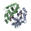

 Mass: 392.491 Da / Num. of mol.: 2 / Source method: obtained synthetically / Formula: C16H30N3O6S
 Mass: 18.015 Da / Num. of mol.: 721 / Source method: isolated from a natural source / Formula: H2O
 Sample preparation
 Processing